 Add to Chrome
 Add to Firefox
 Add to Edge
Automated classification method of COVID-19 cases from chest CT volumes using 2D and 3D hybrid CNN for anisotropic volumes
Jul 31, 2026
This paper proposes an automated classification method of chest CT volumes based on likelihood of COVID-19 cases. Novel coronavirus disease 2019 (COVID-19) spreads over the world, causing a large number of infected patients and deaths. Sudden increase in the number of COVID-19 patients causes a manpower shortage in medical institutions. Computer-aided diagnosis (CAD) system provides quick and quantitative diagnosis results. CAD system for COVID-19 enables efficient diagnosis workflow and contributes to reduce such manpower shortage. This paper proposes an automated classification method of chest CT volumes for COVID-19 diagnosis assistance. We propose a COVID-19 classification convolutional neural network (CNN) that has a 2D/3D hybrid feature extraction flows. The 2D/3D hybrid feature extraction flows are designed to effectively extract image features from anisotropic volumes such as chest CT volumes for diagnosis. The flows extract image features on three mutually perpendicular planes in CT volumes and then combine the features to perform classification. Classification accuracy of the proposed method was evaluated using a dataset that contains 1288 CT volumes. An averaged classification accuracy was 83.3%. The accuracy was higher than that of a classification CNN which does not have 2D and 3D hybrid feature extraction flows.
* Oral Presentation in SPIE Medical Imaging 2022

Classification of COVID-19 cases from chest CT volumes using hybrid model of 3D CNN and 3D MLP-Mixer
Jul 31, 2026This paper proposes an automated classification method of COVID-19 chest CT volumes using improved 3D MLP-Mixer. Novel coronavirus disease 2019 (COVID-19) spreads over the world, causing a large number of infected patients and deaths. Sudden increase in the number of COVID-19 patients causes a manpower shortage in medical institutions. Computer-aided diagnosis (CAD) system provides quick and quantitative diagnosis results. CAD system for COVID-19 enables efficient diagnosis workflow and contributes to reduce such manpower shortage. In image-based diagnosis of viral pneumonia cases including COVID-19, both local and global image features are important because viral pneumonia cause many ground glass opacities and consolidations in large areas in the lung. This paper proposes an automated classification method of chest CT volumes for COVID-19 diagnosis assistance. MLP-Mixer is a recent method of image classification using Vision Transformer-like architecture. It performs classification using both local and global image features. To classify 3D CT volumes, we developed a hybrid classification model that consists of both a 3D convolutional neural network (CNN) and a 3D version of the MLP-Mixer. Classification accuracy of the proposed method was evaluated using a dataset that contains 1205 CT volumes and obtained 79.5% of classification accuracy. The accuracy was higher than that of conventional 3D CNN models consists of 3D CNN layers and simple MLP layers.
* Accepted as a poster presentation in SPIE Medical Imaging 2023
Deep Learning Estimation of Sex, Age, Height, and Weight from CT-derived Digitally Reconstructed Radiographs
Jul 21, 2026Purpose: To develop and validate a deep learning ensemble for estimating adult sex, age, height, and weight from coronal digitally reconstructed radiographs (DRRs) generated from diagnostic CT. Materials and Methods: This retrospective study included 128,621 CT examinations from 80,004 adults at nine institutions in Japan. Three multitask models-ConvNeXt-Base, ViT-Base/16, and MaxViT-Base-were fine-tuned using coronal DRRs and combined by weighted averaging. Data were split by institution into training (114,147 examinations; seven institutions), tuning (4,305; one institution), and test (10,169; one institution) sets; generalizability was assessed on two non-Japanese datasets. Accuracy and mean absolute error (MAE) were used to evaluate sex classification and age, height, and weight regression, respectively. Body surface area (BSA)-corrected heart and liver volume trends were compared using true versus estimated height and weight. Results: In the test set (median age, 69.9 years; 4,899 of 10,169 [48.2%] male), overall sex-classification accuracy was 0.997 (95% CI, 0.996-0.998), and MAEs were 3.57 years (3.51-3.63), 2.59 cm (2.54-2.64), and 3.40 kg (3.34-3.47) for age, height, and weight, respectively. In examinations covering the chest through pelvis, accuracy was 1.000, and MAEs were 3.15 years, 2.28 cm, and 3.18 kg, respectively. BSA calculated from estimated values reproduced age-related heart and liver volume trends obtained using true values. On non-Japanese datasets, height error increased but was reduced by continued fine-tuning. Conclusion: The ensemble estimated adult sex, age, height, and weight from CT-derived DRRs, with generally lower errors in examinations with broader anatomical coverage.
Identifying Suspicious Regions of Covid-19 by Abnormality-Sensitive Activation Mapping
Mar 27, 2023This paper presents a fully-automated method for the identification of suspicious regions of a coronavirus disease (COVID-19) on chest CT volumes. One major role of chest CT scanning in COVID-19 diagnoses is identification of an inflammation particular to the disease. This task is generally performed by radiologists through an interpretation of the CT volumes, however, because of the heavy workload, an automatic analysis method using a computer is desired. Most computer-aided diagnosis studies have addressed only a portion of the elements necessary for the identification. In this work, we realize the identification method through a classification task by using a 2.5-dimensional CNN with three-dimensional attention mechanisms. We visualize the suspicious regions by applying a backpropagation based on positive gradients to attention-weighted features. We perform experiments on an in-house dataset and two public datasets to reveal the generalization ability of the proposed method. The proposed architecture achieved AUCs of over 0.900 for all the datasets, and mean sensitivity $0.853 \pm 0.036$ and specificity $0.870 \pm 0.040$. The method can also identify notable lesions pointed out in the radiology report as suspicious regions.
COVID-19 Infection Segmentation from Chest CT Images Based on Scale Uncertainty
Jan 09, 2022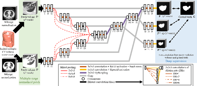

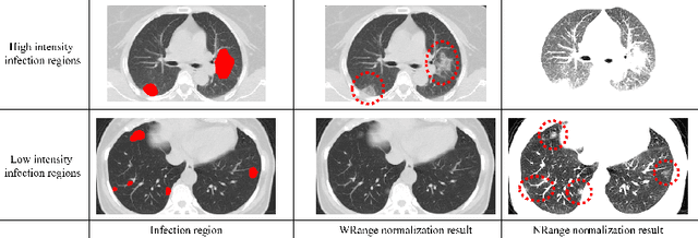

This paper proposes a segmentation method of infection regions in the lung from CT volumes of COVID-19 patients. COVID-19 spread worldwide, causing many infected patients and deaths. CT image-based diagnosis of COVID-19 can provide quick and accurate diagnosis results. An automated segmentation method of infection regions in the lung provides a quantitative criterion for diagnosis. Previous methods employ whole 2D image or 3D volume-based processes. Infection regions have a considerable variation in their sizes. Such processes easily miss small infection regions. Patch-based process is effective for segmenting small targets. However, selecting the appropriate patch size is difficult in infection region segmentation. We utilize the scale uncertainty among various receptive field sizes of a segmentation FCN to obtain infection regions. The receptive field sizes can be defined as the patch size and the resolution of volumes where patches are clipped from. This paper proposes an infection segmentation network (ISNet) that performs patch-based segmentation and a scale uncertainty-aware prediction aggregation method that refines the segmentation result. We design ISNet to segment infection regions that have various intensity values. ISNet has multiple encoding paths to process patch volumes normalized by multiple intensity ranges. We collect prediction results generated by ISNets having various receptive field sizes. Scale uncertainty among the prediction results is extracted by the prediction aggregation method. We use an aggregation FCN to generate a refined segmentation result considering scale uncertainty among the predictions. In our experiments using 199 chest CT volumes of COVID-19 cases, the prediction aggregation method improved the dice similarity score from 47.6% to 62.1%.
* Accepted paper as a oral presentation at CILP2021, 10th MICCAI CLIP Workshop
Lung infection and normal region segmentation from CT volumes of COVID-19 cases
Jan 09, 2022This paper proposes an automated segmentation method of infection and normal regions in the lung from CT volumes of COVID-19 patients. From December 2019, novel coronavirus disease 2019 (COVID-19) spreads over the world and giving significant impacts to our economic activities and daily lives. To diagnose the large number of infected patients, diagnosis assistance by computers is needed. Chest CT is effective for diagnosis of viral pneumonia including COVID-19. A quantitative analysis method of condition of the lung from CT volumes by computers is required for diagnosis assistance of COVID-19. This paper proposes an automated segmentation method of infection and normal regions in the lung from CT volumes using a COVID-19 segmentation fully convolutional network (FCN). In diagnosis of lung diseases including COVID-19, analysis of conditions of normal and infection regions in the lung is important. Our method recognizes and segments lung normal and infection regions in CT volumes. To segment infection regions that have various shapes and sizes, we introduced dense pooling connections and dilated convolutions in our FCN. We applied the proposed method to CT volumes of COVID-19 cases. From mild to severe cases of COVID-19, the proposed method correctly segmented normal and infection regions in the lung. Dice scores of normal and infection regions were 0.911 and 0.753, respectively.
* Accepted paper as a poster presentation at SPIE Medical Imaging 2021