 Add to Chrome
 Add to Firefox
 Add to Edge
On the Adversarial Robustness of Large Vision-Language Models under Visual Token Compression
Jan 29, 2026Visual token compression is widely used to accelerate large vision-language models (LVLMs) by pruning or merging visual tokens, yet its adversarial robustness remains unexplored. We show that existing encoder-based attacks can substantially overestimate the robustness of compressed LVLMs, due to an optimization-inference mismatch: perturbations are optimized on the full-token representation, while inference is performed through a token-compression bottleneck. To address this gap, we propose the Compression-AliGnEd attack (CAGE), which aligns perturbation optimization with compression inference without assuming access to the deployed compression mechanism or its token budget. CAGE combines (i) expected feature disruption, which concentrates distortion on tokens likely to survive across plausible budgets, and (ii) rank distortion alignment, which actively aligns token distortions with rank scores to promote the retention of highly distorted evidence. Across diverse representative plug-and-play compression mechanisms and datasets, our results show that CAGE consistently achieves lower robust accuracy than the baseline. This work highlights that robustness assessments ignoring compression can be overly optimistic, calling for compression-aware security evaluation and defenses for efficient LVLMs.
A Sample-Level Evaluation and Generative Framework for Model Inversion Attacks
Feb 26, 2025Model Inversion (MI) attacks, which reconstruct the training dataset of neural networks, pose significant privacy concerns in machine learning. Recent MI attacks have managed to reconstruct realistic label-level private data, such as the general appearance of a target person from all training images labeled on him. Beyond label-level privacy, in this paper we show sample-level privacy, the private information of a single target sample, is also important but under-explored in the MI literature due to the limitations of existing evaluation metrics. To address this gap, this study introduces a novel metric tailored for training-sample analysis, namely, the Diversity and Distance Composite Score (DDCS), which evaluates the reconstruction fidelity of each training sample by encompassing various MI attack attributes. This, in turn, enhances the precision of sample-level privacy assessments. Leveraging DDCS as a new evaluative lens, we observe that many training samples remain resilient against even the most advanced MI attack. As such, we further propose a transfer learning framework that augments the generative capabilities of MI attackers through the integration of entropy loss and natural gradient descent. Extensive experiments verify the effectiveness of our framework on improving state-of-the-art MI attacks over various metrics including DDCS, coverage and FID. Finally, we demonstrate that DDCS can also be useful for MI defense, by identifying samples susceptible to MI attacks in an unsupervised manner.
A Learning-from-noise Dilated Wide Activation Network for denoising Arterial Spin Labeling Perfusion Images
May 15, 2020
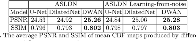
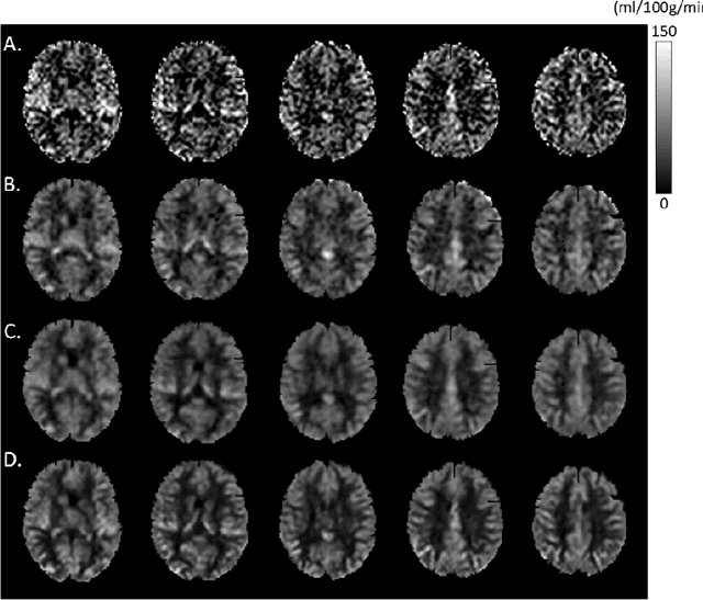
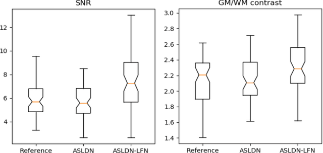
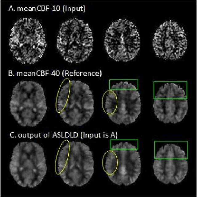
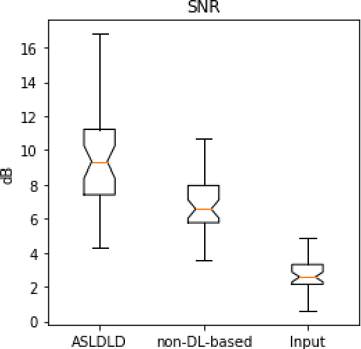
Arterial spin labeling (ASL) perfusion MRI provides a non-invasive way to quantify cerebral blood flow (CBF) but it still suffers from a low signal-to-noise-ratio (SNR). Using deep machine learning (DL), several groups have shown encouraging denoising results. Interestingly, the improvement was obtained when the deep neural network was trained using noise-contaminated surrogate reference because of the lack of golden standard high quality ASL CBF images. More strikingly, the output of these DL ASL networks (ASLDN) showed even higher SNR than the surrogate reference. This phenomenon indicates a learning-from-noise capability of deep networks for ASL CBF image denoising, which can be further enhanced by network optimization. In this study, we proposed a new ASLDN to test whether similar or even better ASL CBF image quality can be achieved in the case of highly noisy training reference. Different experiments were performed to validate the learning-from-noise hypothesis. The results showed that the learning-from-noise strategy produced better output quality than ASLDN trained with relatively high SNR reference.
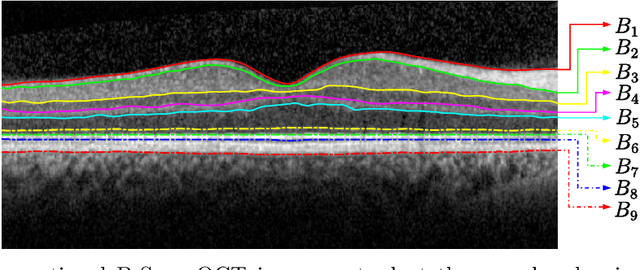
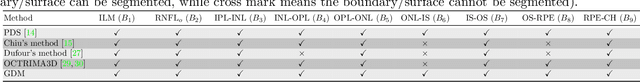
OCT segmentation: Integrating open parametric contour model of the retinal layers and shape constraint to the Mumford-Shah functional
Aug 08, 2018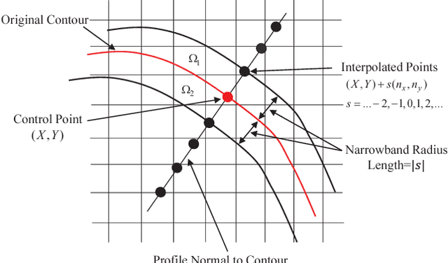
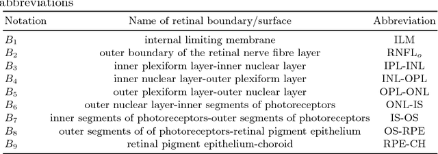
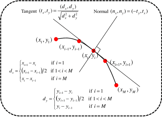
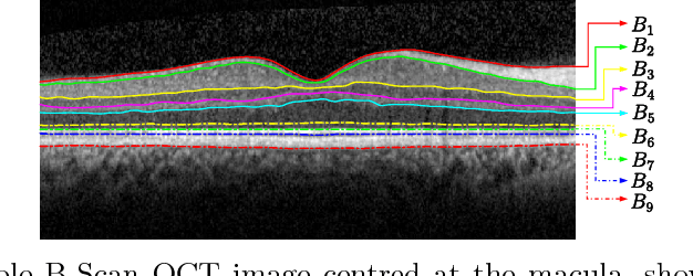
In this paper, we propose a novel retinal layer boundary model for segmentation of optical coherence tomography (OCT) images. The retinal layer boundary model consists of 9 open parametric contours representing the 9 retinal layers in OCT images. An intensity-based Mumford-Shah (MS) variational functional is first defined to evolve the retinal layer boundary model to segment the 9 layers simultaneously. By making use of the normals of open parametric contours, we construct equal sized adjacent narrowbands that are divided by each contour. Regional information in each narrowband can thus be integrated into the MS energy functional such that its optimisation is robust against different initialisations. A statistical prior is also imposed on the shape of the segmented parametric contours for the functional. As such, by minimising the MS energy functional the parametric contours can be driven towards the true boundaries of retinal layers, while the similarity of the contours with respect to training OCT shapes is preserved. Experimental results on real OCT images demonstrate that the method is accurate and robust to low quality OCT images with low contrast and high-level speckle noise, and it outperforms the recent geodesic distance based method for segmenting 9 layers of the retina in OCT images.
Denoising Arterial Spin Labeling Cerebral Blood Flow Images Using Deep Learning
Jan 29, 2018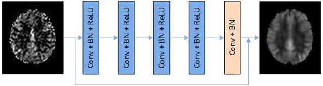
Arterial spin labeling perfusion MRI is a noninvasive technique for measuring quantitative cerebral blood flow (CBF), but the measurement is subject to a low signal-to-noise-ratio(SNR). Various post-processing methods have been proposed to denoise ASL MRI but only provide moderate improvement. Deep learning (DL) is an emerging technique that can learn the most representative signal from data without prior modeling which can be highly complex and analytically indescribable. The purpose of this study was to assess whether the record breaking performance of DL can be translated into ASL MRI denoising. We used convolutional neural network (CNN) to build the DL ASL denosing model (DL-ASL) to inherently consider the inter-voxel correlations. To better guide DL-ASL training, we incorporated prior knowledge about ASL MRI: the structural similarity between ASL CBF map and grey matter probability map. A relatively large sample data were used to train the model which was subsequently applied to a new set of data for testing. Experimental results showed that DL-ASL achieved state-of-the-art denoising performance for ASL MRI as compared to current routine methods in terms of higher SNR, keeping CBF quantification quality while shorten the acquisition time by 75%, and automatic partial volume correction.
Tensor Based Second Order Variational Model for Image Reconstruction
Sep 27, 2016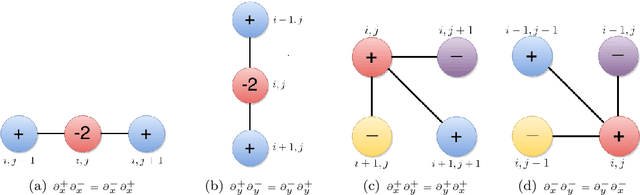
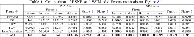
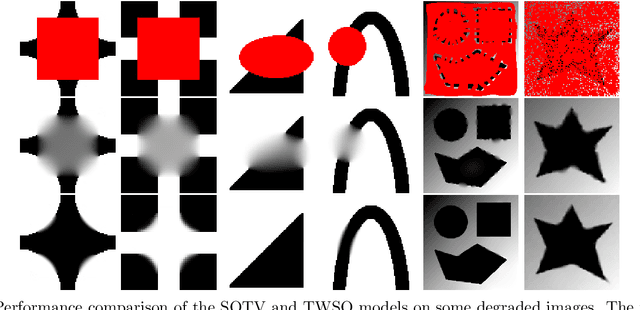
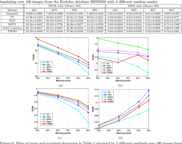
Second order total variation (SOTV) models have advantages for image reconstruction over their first order counterparts including their ability to remove the staircase artefact in the reconstructed image, but they tend to blur the reconstructed image. To overcome this drawback, we introduce a new Tensor Weighted Second Order (TWSO) model for image reconstruction. Specifically, we develop a novel regulariser for the SOTV model that uses the Frobenius norm of the product of the SOTV Hessian matrix and the anisotropic tensor. We then adapt the alternating direction method of multipliers (ADMM) to solve the proposed model by breaking down the original problem into several subproblems. All the subproblems have closed-forms and can thus be solved efficiently. The proposed method is compared with a range of state-of-the-art approaches such as tensor-based anisotropic diffusion, total generalised variation, Euler's elastica, etc. Numerical experimental results of the method on both synthetic and real images from the Berkeley database BSDS500 demonstrate that the proposed method eliminates both the staircase and blurring effects and outperforms the existing approaches for image inpainting and denoising applications.
Automated Segmentation of Retinal Layers from Optical Coherent Tomography Images Using Geodesic Distance
Sep 07, 2016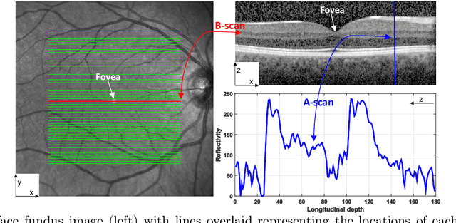
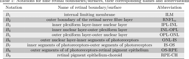
Optical coherence tomography (OCT) is a non-invasive imaging technique that can produce images of the eye at the microscopic level. OCT image segmentation to localise retinal layer boundaries is a fundamental procedure for diagnosing and monitoring the progression of retinal and optical nerve disorders. In this paper, we introduce a novel and accurate geodesic distance method (GDM) for OCT segmentation of both healthy and pathological images in either two- or three-dimensional spaces. The method uses a weighted geodesic distance by an exponential function, taking into account both horizontal and vertical intensity variations. The weighted geodesic distance is efficiently calculated from an Eikonal equation via the fast sweeping method. The segmentation is then realised by solving an ordinary differential equation with the geodesic distance. The results of the GDM are compared with manually segmented retinal layer boundaries/surfaces. Extensive experiments demonstrate that the proposed GDM is robust to complex retinal structures with large curvatures and irregularities and it outperforms the parametric active contour algorithm as well as the graph theoretic based approaches for delineating the retinal layers in both healthy and pathological images.
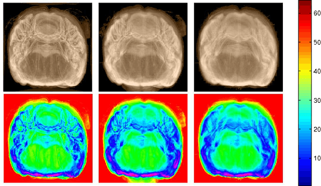
The Influence of Intensity Standardization on Medical Image Registration
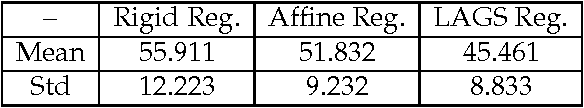
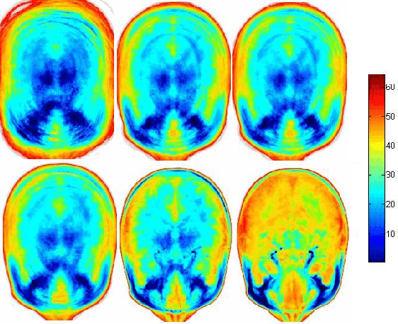
Feb 05, 2010Acquisition-to-acquisition signal intensity variations (non-standardness) are inherent in MR images. Standardization is a post processing method for correcting inter-subject intensity variations through transforming all images from the given image gray scale into a standard gray scale wherein similar intensities achieve similar tissue meanings. The lack of a standard image intensity scale in MRI leads to many difficulties in tissue characterizability, image display, and analysis, including image segmentation. This phenomenon has been documented well; however, effects of standardization on medical image registration have not been studied yet. In this paper, we investigate the influence of intensity standardization in registration tasks with systematic and analytic evaluations involving clinical MR images. We conducted nearly 20,000 clinical MR image registration experiments and evaluated the quality of registrations both quantitatively and qualitatively. The evaluations show that intensity variations between images degrades the accuracy of registration performance. The results imply that the accuracy of image registration not only depends on spatial and geometric similarity but also on the similarity of the intensity values for the same tissues in different images.
Parallel AdaBoost Algorithm for Gabor Wavelet Selection in Face Recognition
Jul 18, 2009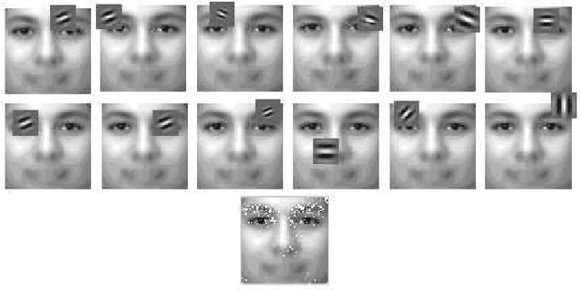
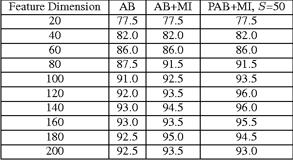
In this paper, the problem of automatic Gabor wavelet selection for face recognition is tackled by introducing an automatic algorithm based on Parallel AdaBoosting method. Incorporating mutual information into the algorithm leads to the selection procedure not only based on classification accuracy but also on efficiency. Effective image features are selected by using properly chosen Gabor wavelets optimised with Parallel AdaBoost method and mutual information to get high recognition rates with low computational cost. Experiments are conducted using the well-known FERET face database. In proposed framework, memory and computation costs are reduced significantly and high classification accuracy is obtained.
* IEEE ICIP 2008 Submission
Fully Automatic 3D Reconstruction of Histological Images
Jul 18, 2009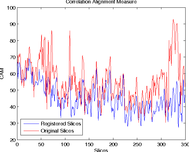
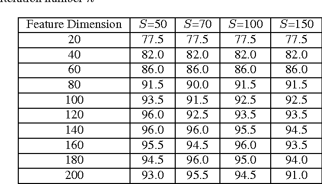
In this paper, we propose a computational framework for 3D volume reconstruction from 2D histological slices using registration algorithms in feature space. To improve the quality of reconstructed 3D volume, first, intensity variations in images are corrected by an intensity standardization process which maps image intensity scale to a standard scale where similar intensities correspond to similar tissues. Second, a subvolume approach is proposed for 3D reconstruction by dividing standardized slices into groups. Third, in order to improve the quality of the reconstruction process, an automatic best reference slice selection algorithm is developed based on an iterative assessment of image entropy and mean square error of the registration process. Finally, we demonstrate that the choice of the reference slice has a significant impact on registration quality and subsequent 3D reconstruction.
* IEEE ISBI-08 Submission