 Add to Chrome
 Add to Firefox
 Add to Edge
DSCA: A Digital Subtraction Angiography Sequence Dataset and Spatio-Temporal Model for Cerebral Artery Segmentation
Jun 01, 2024


Cerebrovascular diseases (CVDs) remain a leading cause of global disability and mortality. Digital Subtraction Angiography (DSA) sequences, recognized as the golden standard for diagnosing CVDs, can clearly visualize the dynamic flow and reveal pathological conditions within the cerebrovasculature. Therefore, precise segmentation of cerebral arteries (CAs) and classification between their main trunks and branches are crucial for physicians to accurately quantify diseases. However, achieving accurate CA segmentation in DSA sequences remains a challenging task due to small vessels with low contrast, and ambiguity between vessels and residual skull structures. Moreover, the lack of publicly available datasets limits exploration in the field. In this paper, we introduce a DSA Sequence-based Cerebral Artery segmentation dataset (DSCA), the first publicly accessible dataset designed specifically for pixel-level semantic segmentation of CAs. Additionally, we propose DSANet, a spatio-temporal network for CA segmentation in DSA sequences. Unlike existing DSA segmentation methods that focus only on a single frame, the proposed DSANet introduces a separate temporal encoding branch to capture dynamic vessel details across multiple frames. To enhance small vessel segmentation and improve vessel connectivity, we design a novel TemporalFormer module to capture global context and correlations among sequential frames. Furthermore, we develop a Spatio-Temporal Fusion (STF) module to effectively integrate spatial and temporal features from the encoder. Extensive experiments demonstrate that DSANet outperforms other state-of-the-art methods in CA segmentation, achieving a Dice of 0.9033.
CS2-Net: Deep Learning Segmentation of Curvilinear Structures in Medical Imaging
Oct 19, 2020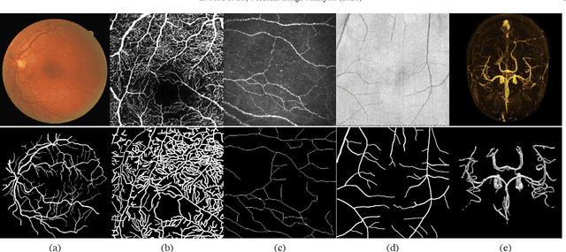
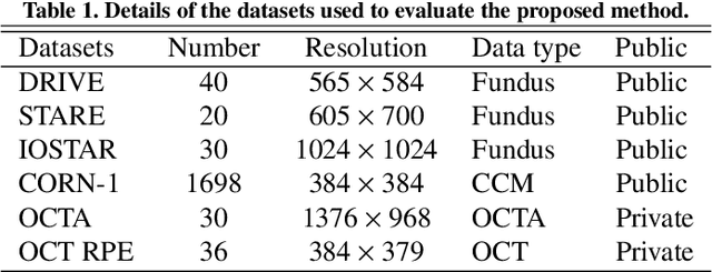
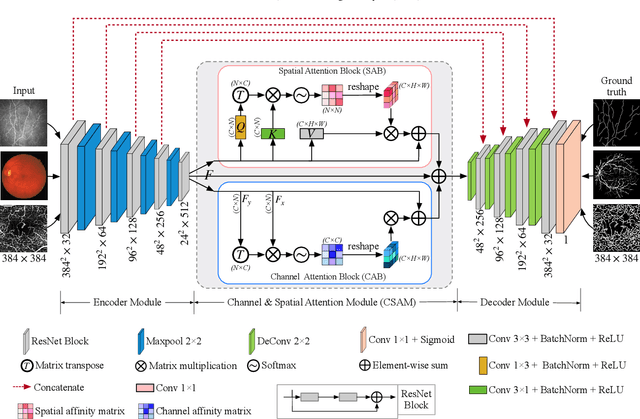
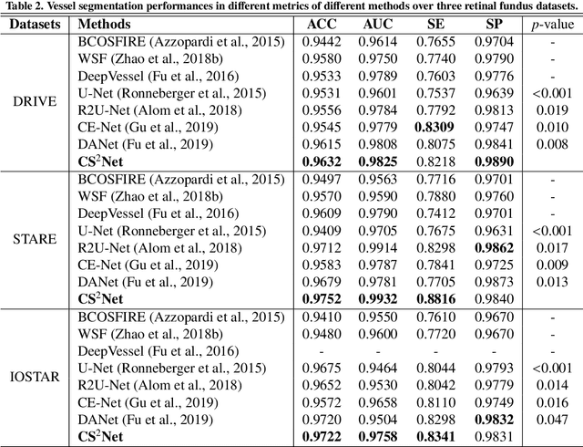
Automated detection of curvilinear structures, e.g., blood vessels or nerve fibres, from medical and biomedical images is a crucial early step in automatic image interpretation associated to the management of many diseases. Precise measurement of the morphological changes of these curvilinear organ structures informs clinicians for understanding the mechanism, diagnosis, and treatment of e.g. cardiovascular, kidney, eye, lung, and neurological conditions. In this work, we propose a generic and unified convolution neural network for the segmentation of curvilinear structures and illustrate in several 2D/3D medical imaging modalities. We introduce a new curvilinear structure segmentation network (CS2-Net), which includes a self-attention mechanism in the encoder and decoder to learn rich hierarchical representations of curvilinear structures. Two types of attention modules - spatial attention and channel attention - are utilized to enhance the inter-class discrimination and intra-class responsiveness, to further integrate local features with their global dependencies and normalization, adaptively. Furthermore, to facilitate the segmentation of curvilinear structures in medical images, we employ a 1x3 and a 3x1 convolutional kernel to capture boundary features. ...
Dense Dilated Network with Probability Regularized Walk for Vessel Detection
Oct 26, 2019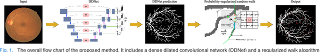
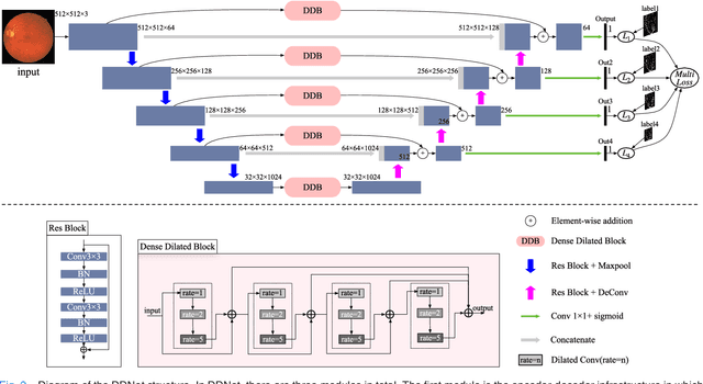
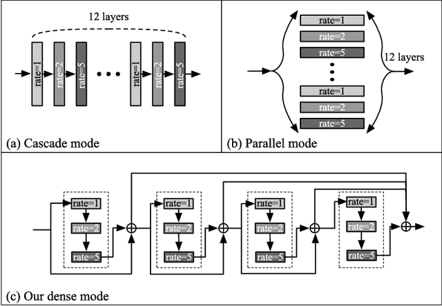
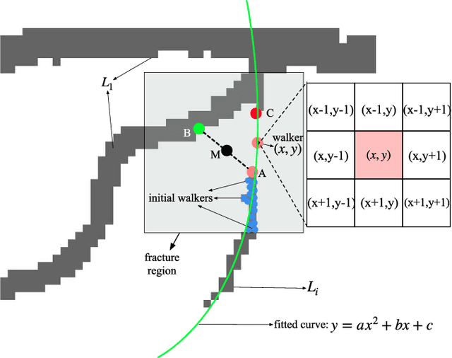
The detection of retinal vessel is of great importance in the diagnosis and treatment of many ocular diseases. Many methods have been proposed for vessel detection. However, most of the algorithms neglect the connectivity of the vessels, which plays an important role in the diagnosis. In this paper, we propose a novel method for retinal vessel detection. The proposed method includes a dense dilated network to get an initial detection of the vessels and a probability regularized walk algorithm to address the fracture issue in the initial detection. The dense dilated network integrates newly proposed dense dilated feature extraction blocks into an encoder-decoder structure to extract and accumulate features at different scales. A multiscale Dice loss function is adopted to train the network. To improve the connectivity of the segmented vessels, we also introduce a probability regularized walk algorithm to connect the broken vessels. The proposed method has been applied on three public data sets: DRIVE, STARE and CHASE_DB1. The results show that the proposed method outperforms the state-of-the-art methods in accuracy, sensitivity, specificity and also are under receiver operating characteristic curve.
The Channel Attention based Context Encoder Network for Inner Limiting Membrane Detection
Aug 09, 2019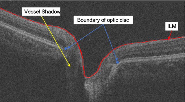
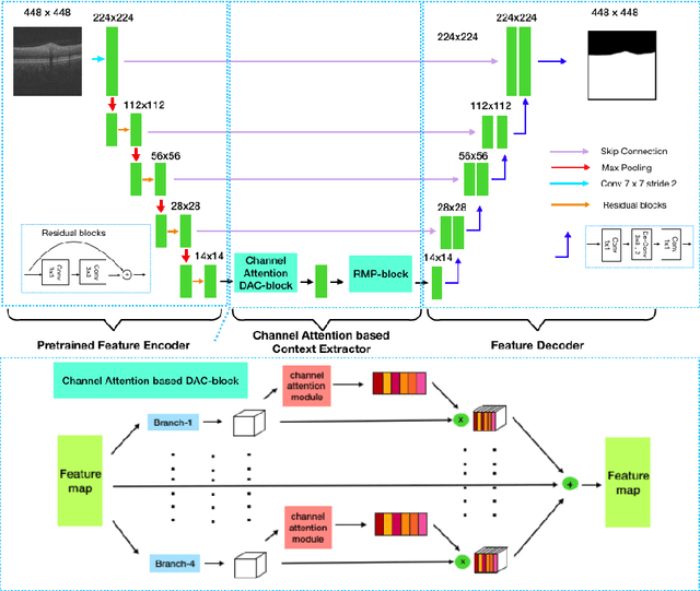
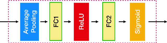
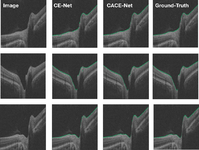
The optic disc segmentation is an important step for retinal image-based disease diagnosis such as glaucoma. The inner limiting membrane (ILM) is the first boundary in the OCT, which can help to extract the retinal pigment epithelium (RPE) through gradient edge information to locate the boundary of the optic disc. Thus, the ILM layer segmentation is of great importance for optic disc localization. In this paper, we build a new optic disc centered dataset from 20 volunteers and manually annotated the ILM boundary in each OCT scan as ground-truth. We also propose a channel attention based context encoder network modified from the CE-Net to segment the optic disc. It mainly contains three phases: the encoder module, the channel attention based context encoder module, and the decoder module. Finally, we demonstrate that our proposed method achieves state-of-the-art disc segmentation performance on our dataset mentioned above.