 Add to Chrome
 Add to Firefox
 Add to Edge
ImmunoDiff: A Diffusion Model for Immunotherapy Response Prediction in Lung Cancer
May 29, 2025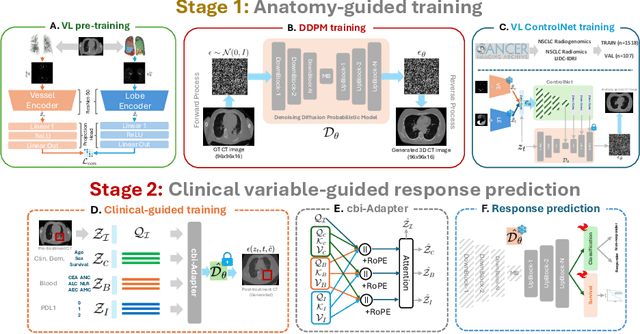

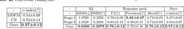

Accurately predicting immunotherapy response in Non-Small Cell Lung Cancer (NSCLC) remains a critical unmet need. Existing radiomics and deep learning-based predictive models rely primarily on pre-treatment imaging to predict categorical response outcomes, limiting their ability to capture the complex morphological and textural transformations induced by immunotherapy. This study introduces ImmunoDiff, an anatomy-aware diffusion model designed to synthesize post-treatment CT scans from baseline imaging while incorporating clinically relevant constraints. The proposed framework integrates anatomical priors, specifically lobar and vascular structures, to enhance fidelity in CT synthesis. Additionally, we introduce a novel cbi-Adapter, a conditioning module that ensures pairwise-consistent multimodal integration of imaging and clinical data embeddings, to refine the generative process. Additionally, a clinical variable conditioning mechanism is introduced, leveraging demographic data, blood-based biomarkers, and PD-L1 expression to refine the generative process. Evaluations on an in-house NSCLC cohort treated with immune checkpoint inhibitors demonstrate a 21.24% improvement in balanced accuracy for response prediction and a 0.03 increase in c-index for survival prediction. Code will be released soon.
Deep-learning-enabled Brain Hemodynamic Mapping Using Resting-state fMRI
Apr 25, 2022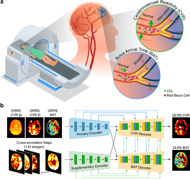
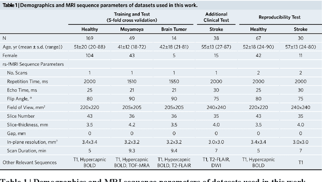
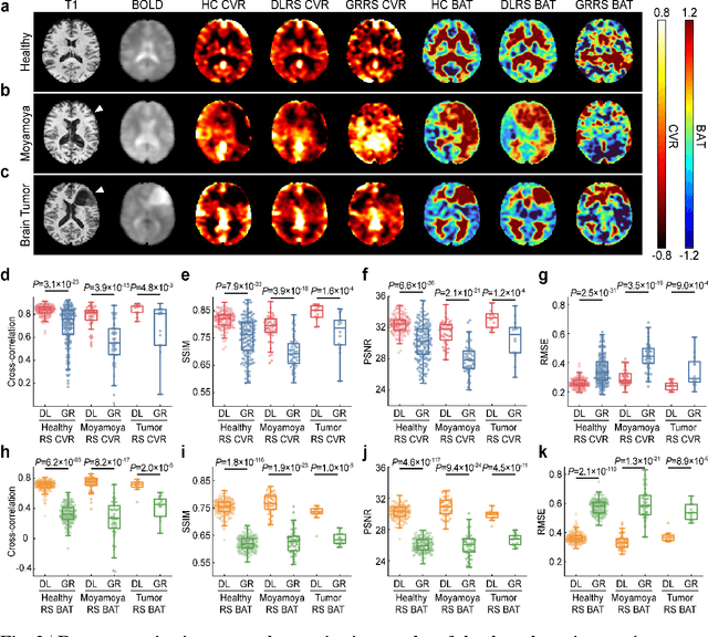
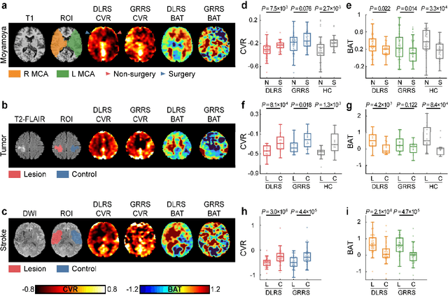
Cerebrovascular disease is a leading cause of death globally. Prevention and early intervention are known to be the most effective forms of its management. Non-invasive imaging methods hold great promises for early stratification, but at present lack the sensitivity for personalized prognosis. Resting-state functional magnetic resonance imaging (rs-fMRI), a powerful tool previously used for mapping neural activity, is available in most hospitals. Here we show that rs-fMRI can be used to map cerebral hemodynamic function and delineate impairment. By exploiting time variations in breathing pattern during rs-fMRI, deep learning enables reproducible mapping of cerebrovascular reactivity (CVR) and bolus arrive time (BAT) of the human brain using resting-state CO2 fluctuations as a natural 'contrast media'. The deep-learning network was trained with CVR and BAT maps obtained with a reference method of CO2-inhalation MRI, which included data from young and older healthy subjects and patients with Moyamoya disease and brain tumors. We demonstrate the performance of deep-learning cerebrovascular mapping in the detection of vascular abnormalities, evaluation of revascularization effects, and vascular alterations in normal aging. In addition, cerebrovascular maps obtained with the proposed method exhibited excellent reproducibility in both healthy volunteers and stroke patients. Deep-learning resting-state vascular imaging has the potential to become a useful tool in clinical cerebrovascular imaging.