 Add to Chrome
 Add to Firefox
 Add to Edge
MR-Compass: Inertial Navigation-Driven Motion Correction for Brain MRI
Mar 02, 2026Inertial sensors can track object kinematics, however, unbounded drift from integrating noisy signals makes them impractical for MRI motion correction at millimeter resolution and minute-long scans. We introduce MR-Compass, which exploits the MRI system's static magnetic and gravitational fields to estimate 3-DOF orientation at 2 kHz directly, without integration, eliminating random-walk. The remaining 3-DOF translation is recovered via phase correlation from the MRI data. We experimentally validate the efficacy of the method retrospectively using a 3D radial koosh-ball sequence and prospectively using 2D EPI fMRI during large volunteer motions. MR-Compass followed by phase-correlation achieved a mean accuracy of 0.6$^o$ and 0.4 pixels across all experiments. Image quality improved when motion correction was applied in all volunteer scans for both retrospective and prospective correction cases. MR-Compass was effective in measuring head motion in the MRI scanner with high accuracy at unprecedented sample rates, and enabled both retrospective and prospective reconstruction to improve image quality by aligning the k-space data appropriately and by reducing the motion related artifacts.
Deep-learning-enabled Brain Hemodynamic Mapping Using Resting-state fMRI
Apr 25, 2022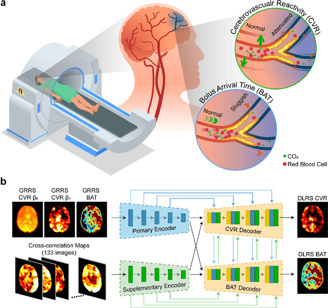
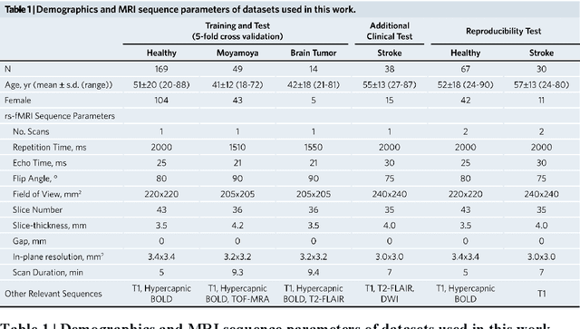
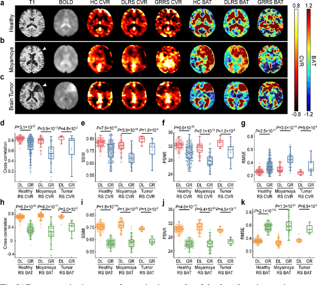
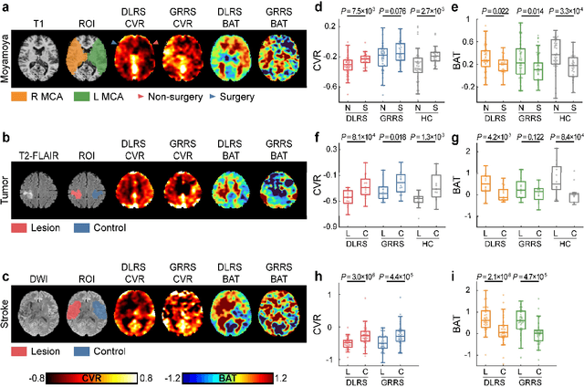
Cerebrovascular disease is a leading cause of death globally. Prevention and early intervention are known to be the most effective forms of its management. Non-invasive imaging methods hold great promises for early stratification, but at present lack the sensitivity for personalized prognosis. Resting-state functional magnetic resonance imaging (rs-fMRI), a powerful tool previously used for mapping neural activity, is available in most hospitals. Here we show that rs-fMRI can be used to map cerebral hemodynamic function and delineate impairment. By exploiting time variations in breathing pattern during rs-fMRI, deep learning enables reproducible mapping of cerebrovascular reactivity (CVR) and bolus arrive time (BAT) of the human brain using resting-state CO2 fluctuations as a natural 'contrast media'. The deep-learning network was trained with CVR and BAT maps obtained with a reference method of CO2-inhalation MRI, which included data from young and older healthy subjects and patients with Moyamoya disease and brain tumors. We demonstrate the performance of deep-learning cerebrovascular mapping in the detection of vascular abnormalities, evaluation of revascularization effects, and vascular alterations in normal aging. In addition, cerebrovascular maps obtained with the proposed method exhibited excellent reproducibility in both healthy volunteers and stroke patients. Deep-learning resting-state vascular imaging has the potential to become a useful tool in clinical cerebrovascular imaging.