 Add to Chrome
 Add to Firefox
 Add to Edge
RetiWave-Mamba: A Dual-Stream Network for Retinal Disease Detection based on Multi-scale Context and Frequency-Adaptive Mamba Projection
Aug 18, 2026
Retinal diseases are a leading cause of irreversible vision impairment, making early and accurate diagnosis essential for effective treatment. Optical Coherence Tomography (OCT) serves as a critical imaging modality for this purpose, yet its automated analysis is hindered by inherent speckle noise, varying lesion scales, and subtle inter-class similarities. To address these challenges, we propose a novel framework, RetiWave-Mamba, which integrates spatial-frequency domain learning with state-of-the-art state space models. The framework utilizes Discrete Wavelet Transform (DWT) to decompose OCT images into low- and high-frequency streams, enabling decoupled processing of structural context and fine-grained details. For the low-frequency branch, we design a Multi-scale Contextual Localization Module (MCLM), which synergizes multi-scale dilation with spatial attention to expand the global receptive field and precisely localize lesion regions. For the high-frequency branch, we introduce an Attention-Guided High-Resolution Network (AG-HRNet) equipped with an intelligent gating mechanism to suppress noise propagation during multi-scale interactions. Furthermore, a Frequency-Adaptive Mamba Projector (FAMP) is incorporated to capture long-range dependencies within disjoint high-frequency textural features. Extensive experiments on the OCT-C8 dataset demonstrate that our approach achieves a state-of-the-art (SOTA) classification accuracy of 98.25%, surpassing existing methods. These results highlight the efficacy of RetiWave-Mamba in robustly identifying retinal pathologies under noisy conditions, offering a promising tool for clinical diagnosis.

$C^3$ASD: Multi-Level Consistency-Driven Representation Learning
Jul 03, 2026Active Speaker Detection determines whether a visible person in a video is speaking at each moment. While recent audio-visual fusion methods perform well on clean data, they degrade under real-world corruptions such as background noise, occlusion, or simultaneous modality degradation. We attribute this limitation to the absence of explicit consistency constraints that promote robust, semantically aligned representations across modalities. Without such guidance, models tend to learn fragile modality-specific shortcuts that fail under corrupted conditions. We propose $C^3$ASD, a multi-level consistency-driven framework with three complementary constraints: embedding-level inter-modality consistency aligns audio-visual representations during speech; sequence-level intra-modality consistency separates speaking and non-speaking clusters via track-aware contrastive learning; and prediction-level consistency stabilizes fusion through knowledge distillation. Extensive experiments demonstrate significant improvements under diverse audio, visual and joint corruptions, while maintaining competitive performance on clean data.
FSS-Net: Frequency-Spatial Synergy Network with Wavelet Attention for Carotid Artery Ultrasound Segmentation
Jun 09, 2026Accurate segmentation of carotid arteries in ultrasound imaging is critical for stroke risk assessment. However, speckle noise, low contrast, and blurred boundaries remain major challenges. In this paper, we propose a Frequency-Spatial Synergy Network (FSS-Net) to achieve noise-robust and high-precision carotid artery segmentation. The network integrates wavelet transform, multi-domain attention, and edge enhancement into a unified encoder-decoder architecture. Specifically, a Channel-Spatial-Wavelet Attention (CSWA) module is designed to suppress noise and purify semantic features in the frequency domain. A Wavelet-Enhanced Bottleneck (WEB) module is introduced to capture long-range global dependencies efficiently. Furthermore, a Laplacian-Guided Adaptive Edge Fusion (LAEF) module compensates high-frequency details and maintains boundary continuity. Extensive experiments on carotid ultrasound datasets show that FSS-Net achieves a Dice score (DSC) of 96.46% and strong robustness under low SNR conditions, outperforming several state-of-the-art methods. This method realizes accurate segmentation of carotid artery in ultrasonic imaging, effectively identifies carotid atherosclerotic plaque, and is verified by other task (such as segmentation of breast cancer), suggesting that it has good clinical application potential in identifying abnormal tissue masses in ultrasonic images.
ClinReadNet: A clinical reading-inspired network for low-dose abdominal CT image quality assessment
Jun 09, 2026In abdominal CT imaging, developing a low-dose, no-reference image quality assessment (No-reference IQA) model that mimics doctors' reading habits for evaluating CT image quality has significant practical value. This paper proposes a novel deep learning-based framework, ClinReadNet, whose design aligns with the clinical reading logic of radiologists: first, it introduces the Sobel ordinal quality network (SOQN) module, which can simultaneously focus on edge details highly relevant to image quality and the quality distribution pattern of the entire image, accurately matching the clinical image-reading judgment habit of "considering both local details and overall context"; second, the framework integrates the (shifted) window multi-scale temperature multi-head self-attention ((S)W-MTMSA) module, which further replicates the radiologists' image-reading process of shifting from overall scanning to local focusing, and accurately locks in regions of interest through multi-sharpness attention; third, it designs the hierarchical ranked probability score (HRPS) loss function, which combines the dual logics of coarse classification and fine classification, while paying attention to the distance information between grading labels, effectively improving the performance of image quality assessment. Experiments conducted on the LDCTIQAG2023 dataset show that the proposed method achieves the current state-of-the-art (SOTA) performance: the values of Pearson's linear correlation coefficient (PLCC), Spearman's rank-order correlation coefficient (SROCC), and Kendall's rank-order correlation coefficient (KROCC) reach 0.9507, 0.9554, and 0.8629 respectively, with the sum of their absolute values (Score) being 2.7690, outperforming existing methods.
CausalDisenSeg: A Causality-Guided Disentanglement Framework with Counterfactual Reasoning for Robust Brain Tumor Segmentation Under Missing Modalities
Apr 15, 2026In clinical practice, the robustness of deep learning models for multimodal brain tumor segmentation is severely compromised by incomplete MRI data. This vulnerability stems primarily from modality bias, where models exploit spurious correlations as shortcuts rather than learning true anatomical structures. Existing feature fusion methods fail to fundamentally eliminate this dependency. To address this, we propose CausalDisenSeg, a novel Structural Causal Model (SCM)-grounded framework that achieves robust segmentation via causality-guided disentanglement and counterfactual reasoning. We reframe the problem as isolating the anatomical Causal Factor from the stylistic Bias Factor. Our framework implements a three-stage causal intervention: (1) Explicit Causal Disentanglement: A Conditional Variational Autoencoder (CVAE) coupled with an HSIC constraint mathematically enforces statistical orthogonality between anatomical and style features. (2) Causal Representation Reinforcement: A Region Causality Module (RCM) explicitly grounds causal features in physical tumor regions. (3) Counterfactual Reasoning: A dual-adversarial strategy actively suppresses the residual Natural Direct Effect (NDE) of the bias, forcing its spatial attention to be mutually exclusive from the causal path. Extensive experiments on the BraTS 2020 dataset demonstrate that CausalDisenSeg significantly outperforms state-of-the-art methods in accuracy and consistency across severe missing-modality scenarios. Furthermore, cross-dataset evaluation on BraTS 2023 under the same protocol yields a state-of-the-art macro-average DSC of 84.49.
MGML: A Plug-and-Play Meta-Guided Multi-Modal Learning Framework for Incomplete Multimodal Brain Tumor Segmentation
Dec 30, 2025Leveraging multimodal information from Magnetic Resonance Imaging (MRI) plays a vital role in lesion segmentation, especially for brain tumors. However, in clinical practice, multimodal MRI data are often incomplete, making it challenging to fully utilize the available information. Therefore, maximizing the utilization of this incomplete multimodal information presents a crucial research challenge. We present a novel meta-guided multi-modal learning (MGML) framework that comprises two components: meta-parameterized adaptive modality fusion and consistency regularization module. The meta-parameterized adaptive modality fusion (Meta-AMF) enables the model to effectively integrate information from multiple modalities under varying input conditions. By generating adaptive soft-label supervision signals based on the available modalities, Meta-AMF explicitly promotes more coherent multimodal fusion. In addition, the consistency regularization module enhances segmentation performance and implicitly reinforces the robustness and generalization of the overall framework. Notably, our approach does not alter the original model architecture and can be conveniently integrated into the training pipeline for end-to-end model optimization. We conducted extensive experiments on the public BraTS2020 and BraTS2023 datasets. Compared to multiple state-of-the-art methods from previous years, our method achieved superior performance. On BraTS2020, for the average Dice scores across fifteen missing modality combinations, building upon the baseline, our method obtained scores of 87.55, 79.36, and 62.67 for the whole tumor (WT), the tumor core (TC), and the enhancing tumor (ET), respectively. We have made our source code publicly available at https://github.com/worldlikerr/MGML.
ERANet: Edge Replacement Augmentation for Semi-Supervised Meniscus Segmentation with Prototype Consistency Alignment and Conditional Self-Training
Feb 11, 2025Manual segmentation is labor-intensive, and automatic segmentation remains challenging due to the inherent variability in meniscal morphology, partial volume effects, and low contrast between the meniscus and surrounding tissues. To address these challenges, we propose ERANet, an innovative semi-supervised framework for meniscus segmentation that effectively leverages both labeled and unlabeled images through advanced augmentation and learning strategies. ERANet integrates three key components: edge replacement augmentation (ERA), prototype consistency alignment (PCA), and a conditional self-training (CST) strategy within a mean teacher architecture. ERA introduces anatomically relevant perturbations by simulating meniscal variations, ensuring that augmentations align with the structural context. PCA enhances segmentation performance by aligning intra-class features and promoting compact, discriminative feature representations, particularly in scenarios with limited labeled data. CST improves segmentation robustness by iteratively refining pseudo-labels and mitigating the impact of label noise during training. Together, these innovations establish ERANet as a robust and scalable solution for meniscus segmentation, effectively addressing key barriers to practical implementation. We validated ERANet comprehensively on 3D Double Echo Steady State (DESS) and 3D Fast/Turbo Spin Echo (FSE/TSE) MRI sequences. The results demonstrate the superior performance of ERANet compared to state-of-the-art methods. The proposed framework achieves reliable and accurate segmentation of meniscus structures, even when trained on minimal labeled data. Extensive ablation studies further highlight the synergistic contributions of ERA, PCA, and CST, solidifying ERANet as a transformative solution for semi-supervised meniscus segmentation in medical imaging.
WaveNet-SF: A Hybrid Network for Retinal Disease Detection Based on Wavelet Transform in the Spatial-Frequency Domain
Jan 21, 2025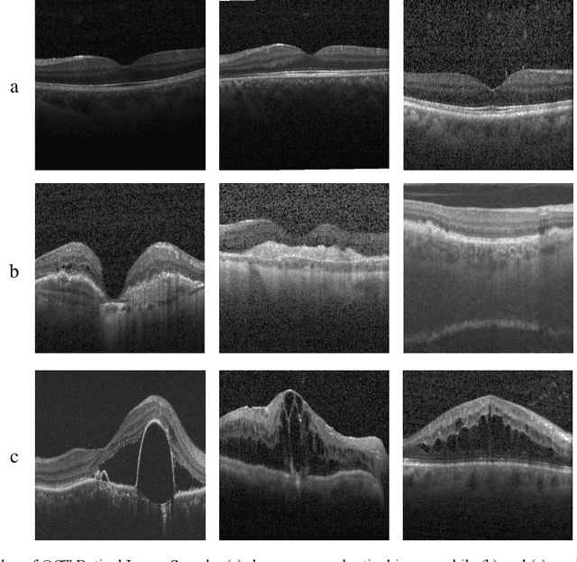
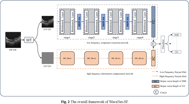
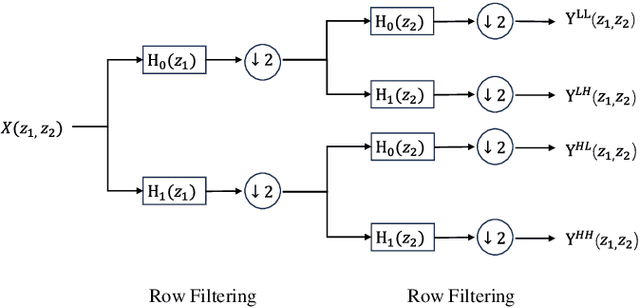
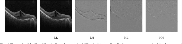
Retinal diseases are a leading cause of vision impairment and blindness, with timely diagnosis being critical for effective treatment. Optical Coherence Tomography (OCT) has become a standard imaging modality for retinal disease diagnosis, but OCT images often suffer from issues such as speckle noise, complex lesion shapes, and varying lesion sizes, making interpretation challenging. In this paper, we propose a novel framework, WaveNet-SF, to enhance retinal disease detection by integrating spatial-domain and frequency-domain learning. The framework utilizes wavelet transforms to decompose OCT images into low- and high-frequency components, enabling the model to extract both global structural features and fine-grained details. To improve lesion detection, we introduce a multi-scale wavelet spatial attention (MSW-SA) module, which enhances the model's focus on regions of interest at multiple scales. Additionally, a high-frequency feature compensation block (HFFC) is incorporated to recover edge information lost during wavelet decomposition, suppress noise, and preserve fine details crucial for lesion detection. Our approach achieves state-of-the-art (SOTA) classification accuracies of 97.82% and 99. 58% on the OCT-C8 and OCT2017 datasets, respectively, surpassing existing methods. These results demonstrate the efficacy of WaveNet-SF in addressing the challenges of OCT image analysis and its potential as a powerful tool for retinal disease diagnosis.
Rethinking domain generalization in medical image segmentation: One image as one domain
Jan 08, 2025Domain shifts in medical image segmentation, particularly when data comes from different centers, pose significant challenges. Intra-center variability, such as differences in scanner models or imaging protocols, can cause domain shifts as large as, or even larger than, those between centers. To address this, we propose the "one image as one domain" (OIOD) hypothesis, which treats each image as a unique domain, enabling flexible and robust domain generalization. Based on this hypothesis, we develop a unified disentanglement-based domain generalization (UniDDG) framework, which simultaneously handles both multi-source and single-source domain generalization without requiring explicit domain labels. This approach simplifies training with a fixed architecture, independent of the number of source domains, reducing complexity and enhancing scalability. We decouple each input image into content representation and style code, then exchange and combine these within the batch for segmentation, reconstruction, and further disentanglement. By maintaining distinct style codes for each image, our model ensures thorough decoupling of content representations and style codes, improving domain invariance of the content representations. Additionally, we enhance generalization with expansion mask attention (EMA) for boundary preservation and style augmentation (SA) to simulate diverse image styles, improving robustness to domain shifts. Extensive experiments show that our method achieves Dice scores of 84.43% and 88.91% for multi-source to single-center and single-center generalization in optic disc and optic cup segmentation, respectively, and 86.96% and 88.56% for prostate segmentation, outperforming current state-of-the-art domain generalization methods, offering superior performance and adaptability across clinical settings.
DGSSA: Domain generalization with structural and stylistic augmentation for retinal vessel segmentation
Jan 07, 2025

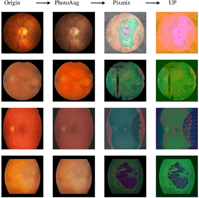
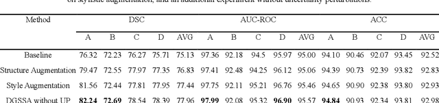
Retinal vascular morphology is crucial for diagnosing diseases such as diabetes, glaucoma, and hypertension, making accurate segmentation of retinal vessels essential for early intervention. Traditional segmentation methods assume that training and testing data share similar distributions, which can lead to poor performance on unseen domains due to domain shifts caused by variations in imaging devices and patient demographics. This paper presents a novel approach, DGSSA, for retinal vessel image segmentation that enhances model generalization by combining structural and style augmentation strategies. We utilize a space colonization algorithm to generate diverse vascular-like structures that closely mimic actual retinal vessels, which are then used to generate pseudo-retinal images with an improved Pix2Pix model, allowing the segmentation model to learn a broader range of structure distributions. Additionally, we utilize PixMix to implement random photometric augmentations and introduce uncertainty perturbations, thereby enriching stylistic diversity and significantly enhancing the model's adaptability to varying imaging conditions. Our framework has been rigorously evaluated on four challenging datasets-DRIVE, CHASEDB, HRF, and STARE-demonstrating state-of-the-art performance that surpasses existing methods. This validates the effectiveness of our proposed approach, highlighting its potential for clinical application in automated retinal vessel analysis.