 Add to Chrome
 Add to Firefox
 Add to Edge
Reconstructing Historical Manuscripts through MSI: The Potential of Contrast in Assessing Image Quality and Legibility
Aug 13, 2026
Digital restoration of historical manuscript images aims to improve readability while preserving the authenticity of cultural heritage documents. However, evaluating quality of restored manuscripts remains challenging, where readability is often subjective and expert annotations are scarce. This study investigates the suitability of contrast-based image quality measures to assess quality and legibility of reconstructed manuscript images from multi-spectral imaging. Two experiments were conducted with publicly-available data sets, facilitating manual quality scores by experts and full-reference image quality measures as reference evaluations. The results show that potential contrast achieves the highest correlation with expert ratings, while contrast-to-noise ratio demonstrates the strongest agreement with full-reference quality measures. Overall, contrast-based measures consistently outperform general image quality measures, demonstrating their potential as objective indicators of manuscript legibility and reconstruction quality.
* 8 pages main paper, 7 pages Supplementary Material

CBCT-IQ: A Publicly Available Annotated Cone-Beam CT Dataset for Image Quality Assessment and Benchmarking
Jul 31, 2026Medical image quality plays a critical role in diagnostic accuracy, especially in X-ray-based imaging modalities such as cone-beam computed tomography (CBCT), where image quality must be balanced against radiation dose. While expert visual evaluation remains the clinical standard for image quality evaluation, it is time-consuming, subjective and affected by inter-observer variability, emphasizing the need for reliable quantitative image quality assessment (IQA) methods. However, the development and validation of such IQA methods have been limited by the lack of publicly available CBCT datasets with expert image quality annotations. In this study, we provide the first open-access CBCT IQA dataset containing 1,764 annotated image slices acquired using systematic variations in image acquisition and reconstruction parameters. Three clinical experts graded the overall image quality and a predefined regions of interest (ROI) using a four-level scoring scheme. In addition, we benchmark 26 full reference- and no reference-based IQA measures against expert annotations and introduce an exploratory IQA measure-based ranking capable of distinguishing subtle image quality differences. This dataset introduced a standardized benchmark for future CBCT IQA research and provides a valuable resource for the development and validation of new IQA methods, enabling reproducible research and advancing CBCT IQA.
Rethinking Clinical Relevance in Chest X-ray Machine Learning: How Evaluation References Define Performance
Jul 28, 2026Chest X-ray (CXR) machine learning relies heavily on automated evaluation using reference standards that aim to approximate clinical judgment. However, commonly used report-derived labels for pathology classification or generic image quality metrics for reconstruction may not reliably reflect clinical judgment. We systematically investigate how evaluation-reference choices affect model performance and ranking in both pathology classification and image quality assessment (IQA). To enable controlled comparison across evaluation references, we collected paired expert image- and report-derived labels for thoracic findings from a clinical cohort at Cambridge University Hospitals (CUH) and curated a subset of the public MIMIC-CXR dataset, along with expert ratings of diagnostic image quality. We show that for supervised image classifiers (ResNet, DenseNet), several zero-shot and fine-tuned vision-language models (e.g., MedKLIP, GLoRIA, and ConVIRT), changing the label source leads to substantial differences not only in performance estimates but also in model rankings. In parallel, alignment of IQA measures with expert judgment depends heavily on the choice of measure, and commonly used IQA metrics such as SSIM and PSNR often fail to align with expert assessments of diagnostic usability. Our results demonstrate that evaluation choices are crucial: they can determine which models and methods appear best and are therefore selected for further development or deployment. The selection of evaluation references should therefore be treated as a central component of clinical validity in CXR machine learning, and justified with respect to the pathology, imaging task, and intended downstream clinical use.
No-reference based automatic parameter optimization for iterative reconstruction using a novel search space aware crow search algorithm
Apr 06, 2026Iterative reconstruction technique's ability to reduce radiation exposure by using fewer projections has attracted significant attention. However, these methods typically require a precise tuning of several hyperparameters, which can have a major impact on reconstruction quality. Manually setting these parameters is time-consuming and increases the workload for human operators. In this paper, we introduce a novel fully automatic parameter optimization framework that can be applied to a wide range of Cone-beam computed tomography (CBCT) iterative reconstruction algorithms to determine optimal parameters without requiring a reference reconstruction. The proposed method incorporates a modified crow search algorithm (CSA) featuring a superior set-dependent local search mechanism, a search-space-aware global search strategy, and an objective-driven balance between local and global search. Additionally, to ensure an effective initial population, we propose a chaotic diagonal linear uniform initialization scheme that accelerates algorithm convergence. The performance of the proposed framework was evaluated on three imaging machines and four real datasets, as well as three different iterative reconstruction methods with the highest number of tunable parameters, representing the most challenging senario. The results indicate that the proposed method could outperform manual settings and CSA, with an 4.19% improvement in average fitness and 4.89% and 3.82% improvements on CHILL@UK and RPI_AXIS, respectively, which are two benchmark no-reference learning-based quality metrics. In addition, the qualitative results clearly show the superiority of the proposed method by maintaining fine details sharply. The overall performance of the proposed framework across different comparison scenarios demonstrates its effectiveness and robustness across all cases.
Potential Contrast: Properties, Equivalences, and Generalization to Multiple Classes
May 02, 2025Potential contrast is typically used as an image quality measure and quantifies the maximal possible contrast between samples from two classes of pixels in an image after an arbitrary grayscale transformation. It has been valuable in cultural heritage applications, identifying and visualizing relevant information in multispectral images while requiring a small number of pixels to be manually sampled. In this work, we introduce a normalized version of potential contrast that removes dependence on image format and also prove equalities that enable generalization to more than two classes and to continuous settings. Finally, we exemplify the utility of multi-class normalized potential contrast through an application to a medieval music manuscript with visible bleedthrough from the back page. We share our implementations, based on both original algorithms and our new equalities, including generalization to multiple classes, at https://github.com/wallacepeaslee/Multiple-Class-Normalized-Potential-Contrast.
Parameter choices in HaarPSI for IQA with medical images
Oct 31, 2024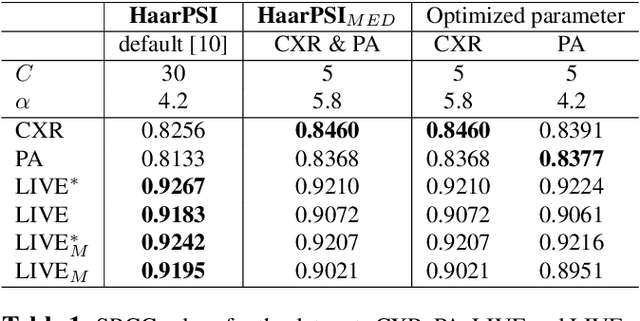
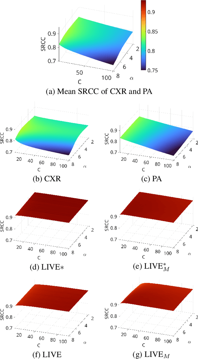
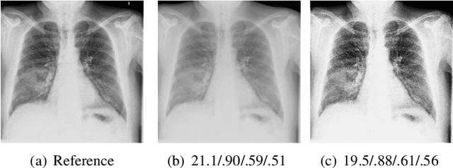
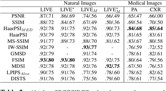
When developing machine learning models, image quality assessment (IQA) measures are a crucial component for evaluation. However, commonly used IQA measures have been primarily developed and optimized for natural images. In many specialized settings, such as medical images, this poses an often-overlooked problem regarding suitability. In previous studies, the IQA measure HaarPSI showed promising behavior for natural and medical images. HaarPSI is based on Haar wavelet representations and the framework allows optimization of two parameters. So far, these parameters have been aligned for natural images. Here, we optimize these parameters for two annotated medical data sets, a photoacoustic and a chest X-Ray data set. We observe that they are more sensitive to the parameter choices than the employed natural images, and on the other hand both medical data sets lead to similar parameter values when optimized. We denote the optimized setting, which improves the performance for the medical images notably, by HaarPSI$_{MED}$. The results suggest that adapting common IQA measures within their frameworks for medical images can provide a valuable, generalizable addition to the employment of more specific task-based measures.
Can Rule-Based Insights Enhance LLMs for Radiology Report Classification? Introducing the RadPrompt Methodology
Aug 07, 2024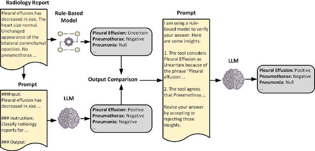
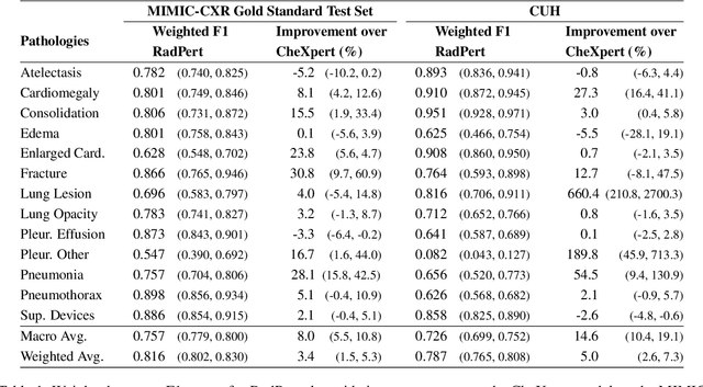
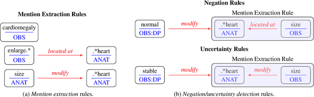
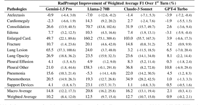
Developing imaging models capable of detecting pathologies from chest X-rays can be cost and time-prohibitive for large datasets as it requires supervision to attain state-of-the-art performance. Instead, labels extracted from radiology reports may serve as distant supervision since these are routinely generated as part of clinical practice. Despite their widespread use, current rule-based methods for label extraction rely on extensive rule sets that are limited in their robustness to syntactic variability. To alleviate these limitations, we introduce RadPert, a rule-based system that integrates an uncertainty-aware information schema with a streamlined set of rules, enhancing performance. Additionally, we have developed RadPrompt, a multi-turn prompting strategy that leverages RadPert to bolster the zero-shot predictive capabilities of large language models, achieving a statistically significant improvement in weighted average F1 score over GPT-4 Turbo. Most notably, RadPrompt surpasses both its underlying models, showcasing the synergistic potential of LLMs with rule-based models. We have evaluated our methods on two English Corpora: the MIMIC-CXR gold-standard test set and a gold-standard dataset collected from the Cambridge University Hospitals.
A study on the adequacy of common IQA measures for medical images
May 29, 2024
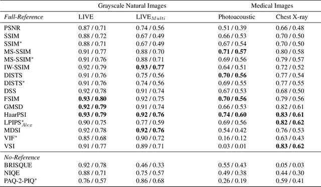


Image quality assessment (IQA) is standard practice in the development stage of novel machine learning algorithms that operate on images. The most commonly used IQA measures have been developed and tested for natural images, but not in the medical setting. Reported inconsistencies arising in medical images are not surprising, as they have different properties than natural images. In this study, we test the applicability of common IQA measures for medical image data by comparing their assessment to manually rated chest X-ray (5 experts) and photoacoustic image data (1 expert). Moreover, we include supplementary studies on grayscale natural images and accelerated brain MRI data. The results of all experiments show a similar outcome in line with previous findings for medical imaging: PSNR and SSIM in the default setting are in the lower range of the result list and HaarPSI outperforms the other tested measures in the overall performance. Also among the top performers in our medical experiments are the full reference measures DISTS, FSIM, LPIPS and MS-SSIM. Generally, the results on natural images yield considerably higher correlations, suggesting that the additional employment of tailored IQA measures for medical imaging algorithms is needed.
A study of why we need to reassess full reference image quality assessment with medical images
May 29, 2024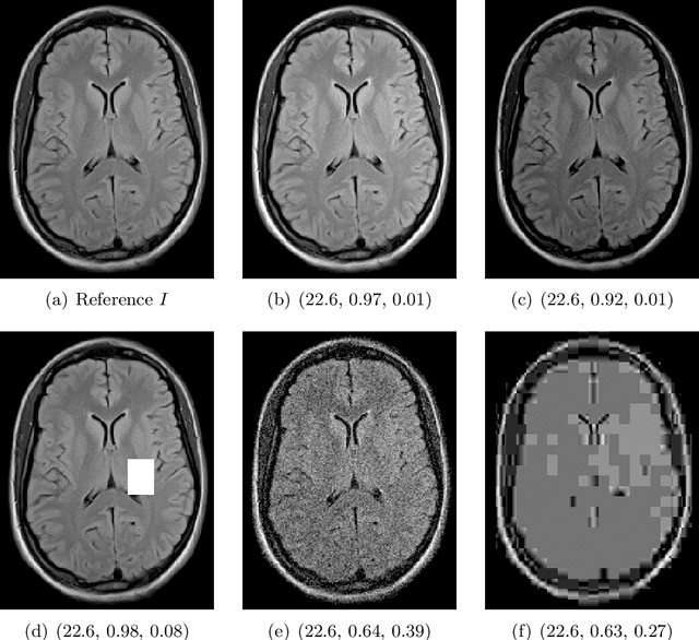
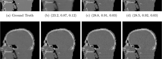
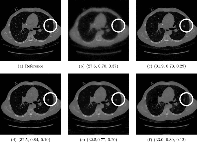
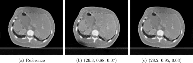
Image quality assessment (IQA) is not just indispensable in clinical practice to ensure high standards, but also in the development stage of novel algorithms that operate on medical images with reference data. This paper provides a structured and comprehensive collection of examples where the two most common full reference (FR) image quality measures prove to be unsuitable for the assessment of novel algorithms using different kinds of medical images, including real-world MRI, CT, OCT, X-Ray, digital pathology and photoacoustic imaging data. In particular, the FR-IQA measures PSNR and SSIM are known and tested for working successfully in many natural imaging tasks, but discrepancies in medical scenarios have been noted in the literature. Inconsistencies arising in medical images are not surprising, as they have very different properties than natural images which have not been targeted nor tested in the development of the mentioned measures, and therefore might imply wrong judgement of novel methods for medical images. Therefore, improvement is urgently needed in particular in this era of AI to increase explainability, reproducibility and generalizability in machine learning for medical imaging and beyond. On top of the pitfalls we will provide ideas for future research as well as suggesting guidelines for the usage of FR-IQA measures applied to medical images.
visClust: A visual clustering algorithm based on orthogonal projections
Nov 07, 2022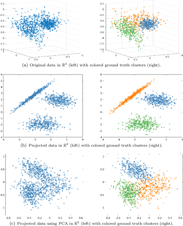

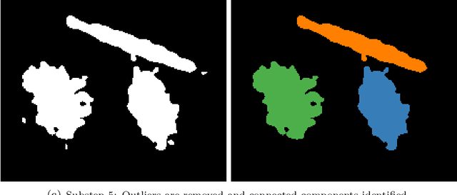
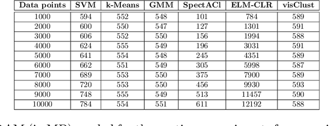
We present a novel clustering algorithm, visClust, that is based on lower dimensional data representations and visual interpretation. Thereto, we design a transformation that allows the data to be represented by a binary integer array enabling the further use of image processing methods to select a partition. Qualitative and quantitative analyses show that the algorithm obtains high accuracy (measured with an adjusted one-sided Rand-Index) and requires low runtime and RAM. We compare the results to 6 state-of-the-art algorithms, confirming the quality of visClust by outperforming in most experiments. Moreover, the algorithm asks for just one obligatory input parameter while allowing optimization via optional parameters. The code is made available on GitHub.