In a research context, image acquisition will often involve a pre-defined static protocol and the data will be of high quality. If we are to build applications that work in hospitals without significant operational changes in care delivery, algorithms should be designed to cope with the available data in the best possible way. In a clinical environment, imaging protocols are highly flexible, with MRI sequences commonly missing appropriate sequence labeling (e.g. T1, T2, FLAIR). To this end we introduce PIMMS, a Permutation Invariant Multi-Modal Segmentation technique that is able to perform inference over sets of MRI scans without using modality labels. We present results which show that our convolutional neural network can, in some settings, outperform a baseline model which utilizes modality labels, and achieve comparable performance otherwise.
Automated medical image segmentation, specifically using deep learning, has shown outstanding performance in semantic segmentation tasks. However, these methods rarely quantify their uncertainty, which may lead to errors in downstream analysis. In this work we propose to use Bayesian neural networks to quantify uncertainty within the domain of semantic segmentation. We also propose a method to convert voxel-wise segmentation uncertainty into volumetric uncertainty, and calibrate the accuracy and reliability of confidence intervals of derived measurements. When applied to a tumour volume estimation application, we demonstrate that by using such modelling of uncertainty, deep learning systems can be made to report volume estimates with well-calibrated error-bars, making them safer for clinical use. We also show that the uncertainty estimates extrapolate to unseen data, and that the confidence intervals are robust in the presence of artificial noise. This could be used to provide a form of quality control and quality assurance, and may permit further adoption of deep learning tools in the clinic.
Multi-task neural network architectures provide a mechanism that jointly integrates information from distinct sources. It is ideal in the context of MR-only radiotherapy planning as it can jointly regress a synthetic CT (synCT) scan and segment organs-at-risk (OAR) from MRI. We propose a probabilistic multi-task network that estimates: 1) intrinsic uncertainty through a heteroscedastic noise model for spatially-adaptive task loss weighting and 2) parameter uncertainty through approximate Bayesian inference. This allows sampling of multiple segmentations and synCTs that share their network representation. We test our model on prostate cancer scans and show that it produces more accurate and consistent synCTs with a better estimation in the variance of the errors, state of the art results in OAR segmentation and a methodology for quality assurance in radiotherapy treatment planning.
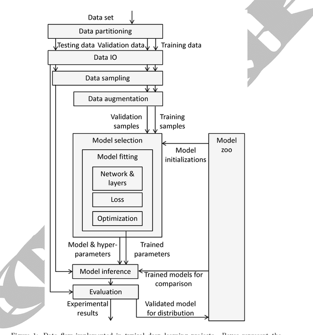
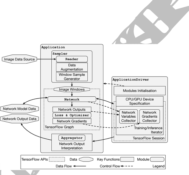
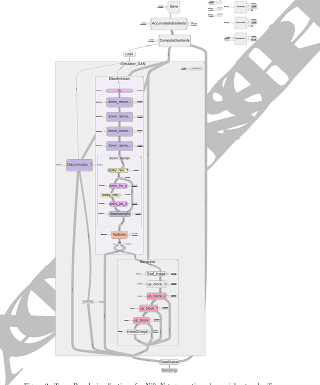
Medical image analysis and computer-assisted intervention problems are increasingly being addressed with deep-learning-based solutions. Established deep-learning platforms are flexible but do not provide specific functionality for medical image analysis and adapting them for this application requires substantial implementation effort. Thus, there has been substantial duplication of effort and incompatible infrastructure developed across many research groups. This work presents the open-source NiftyNet platform for deep learning in medical imaging. The ambition of NiftyNet is to accelerate and simplify the development of these solutions, and to provide a common mechanism for disseminating research outputs for the community to use, adapt and build upon. NiftyNet provides a modular deep-learning pipeline for a range of medical imaging applications including segmentation, regression, image generation and representation learning applications. Components of the NiftyNet pipeline including data loading, data augmentation, network architectures, loss functions and evaluation metrics are tailored to, and take advantage of, the idiosyncracies of medical image analysis and computer-assisted intervention. NiftyNet is built on TensorFlow and supports TensorBoard visualization of 2D and 3D images and computational graphs by default. We present 3 illustrative medical image analysis applications built using NiftyNet: (1) segmentation of multiple abdominal organs from computed tomography; (2) image regression to predict computed tomography attenuation maps from brain magnetic resonance images; and (3) generation of simulated ultrasound images for specified anatomical poses. NiftyNet enables researchers to rapidly develop and distribute deep learning solutions for segmentation, regression, image generation and representation learning applications, or extend the platform to new applications.