 Add to Chrome
 Add to Firefox
 Add to Edge
Human and AI collaboration for pulmonary nodule segmentation
Jun 21, 2026
Medical expert annotators are scarce, and blind reliance on artificial intelligence (AI) can be misleading, motivating approaches in which humans, particularly junior medical trainees or even non-medical personnel, collaborate with AI to achieve robust medical segmentation. Although the Segment Anything Model (SAM) shows promise for general-purpose image segmentation, its performance in human-AI collaboration for specialized medical tasks has not been thoroughly evaluated. Here we present Hi-Seg, a human-in-the-loop segmentation framework for pulmonary nodules built on SAM. Humans iteratively refine prompts through trial-and-error learning and semantic reasoning, progressively guiding SAM toward higher-quality masks. Using chest CT scans from 1,179 patients across 12 centers, we conducted the first large-scale external validation of collaborative human-SAM segmentation. Across all annotator groups, Hi-Seg achieved a mean Dice score of almost 85%, outperforming five state-of-the-art deep learning models by 10-22% and 13 SAM variants by 1-29%. Hi-Seg improved segmentation accuracy while reducing annotation time for medical annotators, and briefly trained non-medical annotators achieved performance comparable to that of the junior medical student. These findings suggest that human-in-the-loop segmentation can reduce clinician workload, enable scalable crowdsourced annotation, and transform clinical workflows by facilitating the safe and efficient integration of foundation models into routine clinical practice.

Alleviating Hyperparameter-Tuning Burden in SVM Classifiers for Pulmonary Nodules Diagnosis with Multi-Task Bayesian Optimization
Nov 09, 2024In the field of non-invasive medical imaging, radiomic features are utilized to measure tumor characteristics. However, these features can be affected by the techniques used to discretize the images, ultimately impacting the accuracy of diagnosis. To investigate the influence of various image discretization methods on diagnosis, it is common practice to evaluate multiple discretization strategies individually. This approach often leads to redundant and time-consuming tasks such as training predictive models and fine-tuning hyperparameters separately. This study examines the feasibility of employing multi-task Bayesian optimization to accelerate the hyperparameters search for classifying benign and malignant pulmonary nodules using RBF SVM. Our findings suggest that multi-task Bayesian optimization significantly accelerates the search for hyperparameters in comparison to a single-task approach. To the best of our knowledge, this is the first investigation to utilize multi-task Bayesian optimization in a critical medical context.
Evolving the pulmonary nodules diagnosis from classical approaches to deep learning aided decision support: three decades development course and future prospect
Jan 23, 2019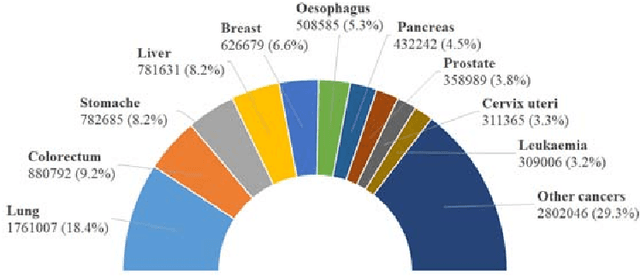
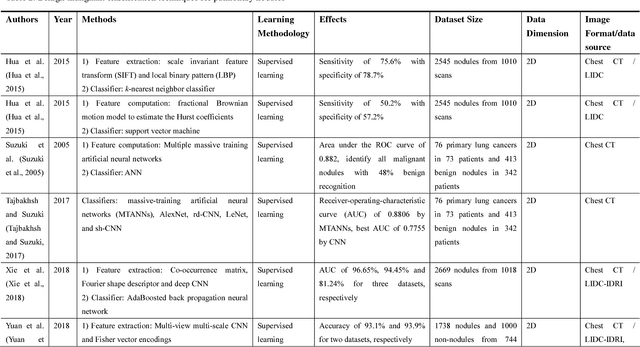
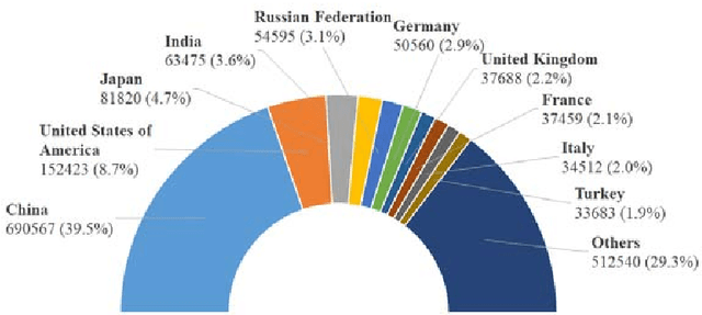
Lung cancer is the commonest cause of cancer deaths worldwide, and its mortality can be reduced significantly by performing early diagnosis and screening. Since the 1960s, driven by the pressing needs to accurately and effectively interpret the massive volume of chest images generated daily, computer-assisted diagnosis of pulmonary nodule has opened up new opportunities to relax the limitation from physicians subjectivity, experiences and fatigue. It has been witnessed that significant and remarkable advances have been achieved since the 1980s, and consistent endeavors have been exerted to deal with the grand challenges on how to accurately detect the pulmonary nodules with high sensitivity at low false-positives rate as well as on how to precisely differentiate between benign and malignant nodules. The main goal of this investigation is to provide a comprehensive state-of-the-art review of the computer-assisted nodules detection and benign-malignant classification techniques developed over three decades, which have evolved from the complicated ad hoc analysis pipeline of conventional approaches to the simplified seamlessly integrated deep learning techniques. This review also identifies challenges and highlights opportunities for future work in learning models, learning algorithms and enhancement schemes for bridging current state to future prospect and satisfying future demand. As far as the authors know, it is the first review of the literature of the past thirty years development in computer-assisted diagnosis of lung nodules. We acknowledge the value of potential multidisciplinary researches that will make the computer-assisted diagnosis of pulmonary nodules enter into the main stream of clinical medicine, and raise the state-of-the-art clinical applications as well as increase both welfares of physicians and patients.