 Add to Chrome
 Add to Firefox
 Add to Edge
Deep Learning Estimation of Sex, Age, Height, and Weight from CT-derived Digitally Reconstructed Radiographs
Jul 21, 2026
Purpose: To develop and validate a deep learning ensemble for estimating adult sex, age, height, and weight from coronal digitally reconstructed radiographs (DRRs) generated from diagnostic CT. Materials and Methods: This retrospective study included 128,621 CT examinations from 80,004 adults at nine institutions in Japan. Three multitask models-ConvNeXt-Base, ViT-Base/16, and MaxViT-Base-were fine-tuned using coronal DRRs and combined by weighted averaging. Data were split by institution into training (114,147 examinations; seven institutions), tuning (4,305; one institution), and test (10,169; one institution) sets; generalizability was assessed on two non-Japanese datasets. Accuracy and mean absolute error (MAE) were used to evaluate sex classification and age, height, and weight regression, respectively. Body surface area (BSA)-corrected heart and liver volume trends were compared using true versus estimated height and weight. Results: In the test set (median age, 69.9 years; 4,899 of 10,169 [48.2%] male), overall sex-classification accuracy was 0.997 (95% CI, 0.996-0.998), and MAEs were 3.57 years (3.51-3.63), 2.59 cm (2.54-2.64), and 3.40 kg (3.34-3.47) for age, height, and weight, respectively. In examinations covering the chest through pelvis, accuracy was 1.000, and MAEs were 3.15 years, 2.28 cm, and 3.18 kg, respectively. BSA calculated from estimated values reproduced age-related heart and liver volume trends obtained using true values. On non-Japanese datasets, height error increased but was reduced by continued fine-tuning. Conclusion: The ensemble estimated adult sex, age, height, and weight from CT-derived DRRs, with generally lower errors in examinations with broader anatomical coverage.

Large Language Model-Assisted Cleaning of Report-Derived Labels in a Large-Scale Chest CT Dataset
Jun 21, 2026Purpose: To evaluate whether large language model (LLM)-assisted label cleaning can identify label-report discordance in CT-RATE, a large-scale public chest CT dataset. Materials and Methods: After report-level deduplication, 24,446 unique radiology reports were identified. Twelve reports were excluded from the primary GPT-5.4 analysis because of Microsoft Azure AI Foundry content-safety filtering, leaving 24,434 reports and 439,812 label instances across 18 abnormality categories. GPT-5.4-derived binary labels were generated from report text using structured JSON output and compared with existing CT-RATE labels. Discordant instances were adjudicated by radiologists. In addition, 100 randomly sampled reports were manually annotated to compare CT-RATE labels, individual LLM-derived labels, and multi-LLM majority-vote labels against radiologist-annotated reference labels. Results: Overall agreement between GPT-5.4-derived and CT-RATE labels was 96.4%, with Cohen's kappa of 0.884. Lymphadenopathy showed the lowest agreement and kappa. In discordance review, radiologist adjudication supported GPT-5.4-derived labels in 72 of 97 (74.2%) general discordant instances and 91 of 99 (91.9%) targeted lymphadenopathy discordant instances. Against radiologist-annotated reference labels, multi-LLM majority-vote labels achieved the highest label-macro-averaged F1 score and Cohen's kappa. Conclusion: LLM-assisted label cleaning identified clinically meaningful label-report discordance in CT-RATE and may support scalable quality improvement of public imaging datasets. The cleaned dataset will be made publicly available to support future research.
Blinded Radiologist and LLM-Based Evaluation of LLM-Generated Japanese Translations of Chest CT Reports: Comparative Study
Apr 02, 2026Background: Accurate translation of radiology reports is important for multilingual research, clinical communication, and radiology education, but the validity of LLM-based evaluation remains unclear. Objective: To evaluate the educational suitability of LLM-generated Japanese translations of chest CT reports and compare radiologist assessments with LLM-as-a-judge evaluations. Methods: We analyzed 150 chest CT reports from the CT-RATE-JPN validation set. For each English report, a human-edited Japanese translation was compared with an LLM-generated translation by DeepSeek-V3.2. A board-certified radiologist and a radiology resident independently performed blinded pairwise evaluations across 4 criteria: terminology accuracy, readability, overall quality, and radiologist-style authenticity. In parallel, 3 LLM judges (DeepSeek-V3.2, Mistral Large 3, and GPT-5) evaluated the same pairs. Agreement was assessed using QWK and percentage agreement. Results: Agreement between radiologists and LLM judges was near zero (QWK=-0.04 to 0.15). Agreement between the 2 radiologists was also poor (QWK=0.01 to 0.06). Radiologist 1 rated terminology as equivalent in 59% of cases and favored the LLM translation for readability (51%) and overall quality (51%). Radiologist 2 rated readability as equivalent in 75% of cases and favored the human-edited translation for overall quality (40% vs 21%). All 3 LLM judges strongly favored the LLM translation across all criteria (70%-99%) and rated it as more radiologist-like in >93% of cases. Conclusions: LLM-generated translations were often judged natural and fluent, but the 2 radiologists differed substantially. LLM-as-a-judge showed strong preference for LLM output and negligible agreement with radiologists. For educational use of translated radiology reports, automated LLM-based evaluation alone is insufficient; expert radiologist review remains important.
ModernBERT is More Efficient than Conventional BERT for Chest CT Findings Classification in Japanese Radiology Reports
Mar 07, 2025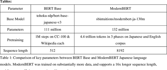

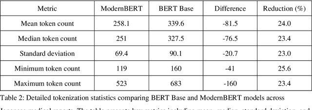

Objective: This study aims to evaluate and compare the performance of two Japanese language models-conventional Bidirectional Encoder Representations from Transformers (BERT) and the newer ModernBERT-in classifying findings from chest CT reports, with a focus on tokenization efficiency, processing time, and classification performance. Methods: We conducted a retrospective study using the CT-RATE-JPN dataset containing 22,778 training reports and 150 test reports. Both models were fine-tuned for multi-label classification of 18 common chest CT conditions. The training data was split in 18,222:4,556 for training and validation. Performance was evaluated using F1 scores for each condition and exact match accuracy across all 18 labels. Results: ModernBERT demonstrated superior tokenization efficiency, requiring 24.0% fewer tokens per document (258.1 vs. 339.6) compared to BERT Base. This translated to significant performance improvements, with ModernBERT completing training in 1877.67 seconds versus BERT's 3090.54 seconds (39% reduction). ModernBERT processed 38.82 samples per second during training (1.65x faster) and 139.90 samples per second during inference (1.66x faster). Despite these efficiency gains, classification performance remained comparable, with ModernBERT achieving superior F1 scores in 8 conditions, while BERT performed better in 4 conditions. Overall exact match accuracy was slightly higher for ModernBERT (74.67% vs. 72.67%), though this difference was not statistically significant (p=0.6291). Conclusion: ModernBERT offers substantial improvements in tokenization efficiency and training speed without sacrificing classification performance. These results suggest that ModernBERT is a promising candidate for clinical applications in Japanese radiology reports analysis.
Development of a Large-scale Dataset of Chest Computed Tomography Reports in Japanese and a High-performance Finding Classification Model
Dec 20, 2024
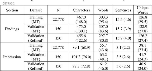


Background: Recent advances in large language models highlight the need for high-quality multilingual medical datasets. While Japan leads globally in CT scanner deployment and utilization, the lack of large-scale Japanese radiology datasets has hindered the development of specialized language models for medical imaging analysis. Objective: To develop a comprehensive Japanese CT report dataset through machine translation and establish a specialized language model for structured finding classification. Additionally, to create a rigorously validated evaluation dataset through expert radiologist review. Methods: We translated the CT-RATE dataset (24,283 CT reports from 21,304 patients) into Japanese using GPT-4o mini. The training dataset consisted of 22,778 machine-translated reports, while the validation dataset included 150 radiologist-revised reports. We developed CT-BERT-JPN based on "tohoku-nlp/bert-base-japanese-v3" architecture for extracting 18 structured findings from Japanese radiology reports. Results: Translation metrics showed strong performance with BLEU scores of 0.731 and 0.690, and ROUGE scores ranging from 0.770 to 0.876 for Findings and from 0.748 to 0.857 for Impression sections. CT-BERT-JPN demonstrated superior performance compared to GPT-4o in 11 out of 18 conditions, including lymphadenopathy (+14.2%), interlobular septal thickening (+10.9%), and atelectasis (+7.4%). The model maintained F1 scores exceeding 0.95 in 14 out of 18 conditions and achieved perfect scores in four conditions. Conclusions: Our study establishes a robust Japanese CT report dataset and demonstrates the effectiveness of a specialized language model for structured finding classification. The hybrid approach of machine translation and expert validation enables the creation of large-scale medical datasets while maintaining high quality.
Zero-shot 3D Segmentation of Abdominal Organs in CT Scans Using Segment Anything Model 2: Adapting Video Tracking Capabilities for 3D Medical Imaging
Aug 12, 2024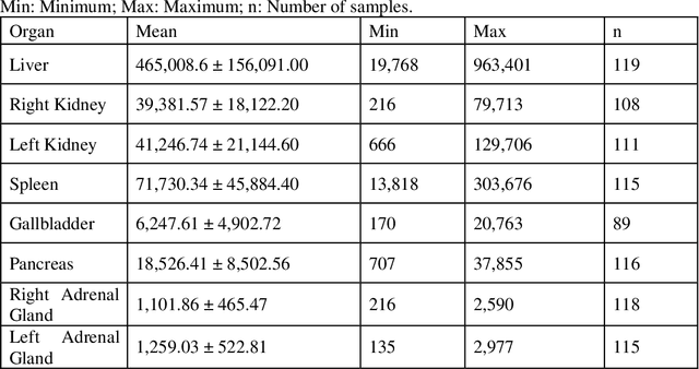
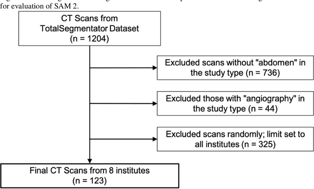
Purpose: This study aimed to evaluate the zero-shot performance of Segment Anything Model 2 (SAM 2) in 3D segmentation of abdominal organs in CT scans, leveraging its video tracking capabilities for volumetric medical imaging. Materials and Methods: Using a subset of the TotalSegmentator CT dataset (n=123) from 8 different institutions, we assessed SAM 2's ability to segment 8 abdominal organs. Segmentation was initiated from three different Z-coordinate levels (caudal, mid, and cranial levels) of each organ. Performance was measured using the Dice similarity coefficient (DSC). We also analyzed organ volumes to contextualize the results. Results: As a zero-shot approach, larger organs with clear boundaries demonstrated high segmentation performance, with mean(median) DSCs as follows: liver 0.821(0.898), left kidney 0.870(0.921), right kidney 0.862(0.935), and spleen 0.891(0.932). Smaller or less defined structures showed lower performance: gallbladder 0.531(0.590), pancreas 0.361(0.359), and adrenal glands 0.203-0.308(0.109-0.231). Significant differences in DSC were observed depending on the starting initial slice of segmentation for different organs. A moderate positive correlation was observed between volume size and DSCs (Spearman's rs = 0.731, P <.001 at caudal-level). DSCs exhibited high variability within organs, ranging from near 0 to almost 1.0, indicating substantial inconsistency in segmentation performance between scans. Conclusion: SAM 2 demonstrated promising zero-shot performance in segmenting certain abdominal organs in CT scans, particularly larger organs with clear boundaries. The model's ability to segment previously unseen targets without additional training highlights its potential for cross-domain generalization in medical imaging. However, improvements are needed for smaller and less defined structures.
Synthetic data generation method for hybrid image-tabular data using two generative adversarial networks
Aug 15, 2023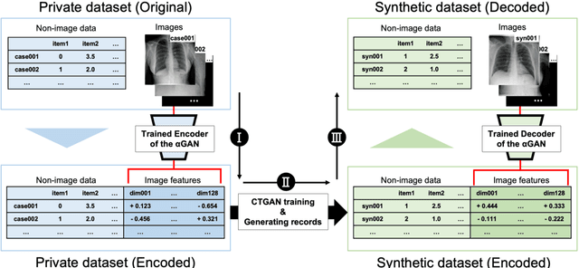

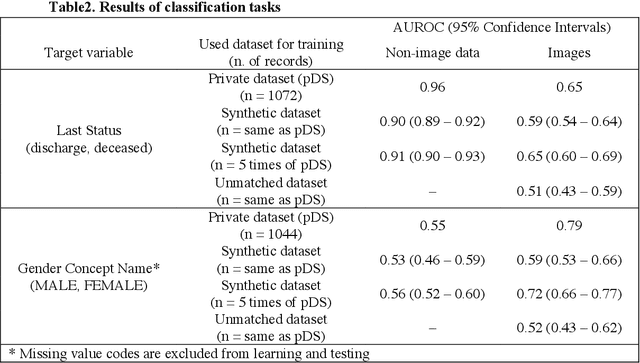
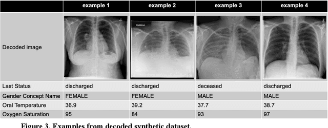
The generation of synthetic medical records using generative adversarial networks (GANs) has become increasingly important for addressing privacy concerns and promoting data sharing in the medical field. In this paper, we propose a novel method for generating synthetic hybrid medical records consisting of chest X-ray images (CXRs) and structured tabular data (including anthropometric data and laboratory tests) using an auto-encoding GAN ({\alpha}GAN) and a conditional tabular GAN (CTGAN). Our approach involves training a {\alpha}GAN model on a large public database (pDB) to reduce the dimensionality of CXRs. We then applied the trained encoder of the GAN model to the images in original database (oDB) to obtain the latent vectors. These latent vectors were combined with tabular data in oDB, and these joint data were used to train the CTGAN model. We successfully generated diverse synthetic records of hybrid CXR and tabular data, maintaining correspondence between them. We evaluated this synthetic database (sDB) through visual assessment, distribution of interrecord distances, and classification tasks. Our evaluation results showed that the sDB captured the features of the oDB while maintaining the correspondence between the images and tabular data. Although our approach relies on the availability of a large-scale pDB containing a substantial number of images with the same modality and imaging region as those in the oDB, this method has the potential for the public release of synthetic datasets without compromising the secondary use of data.