 Add to Chrome
 Add to Firefox
 Add to Edge
Automatic Landmark-Based Segmentation of Human Subcortical Structures in MRI
May 14, 2026Precise segmentation of brain structures in magnetic resonance imaging (MRI) is essential for reliable neuroimaging analysis, yet voxel-wise deep models often yield anatomically inconsistent results that diverge from expert-defined boundaries. In this research, we propose a landmark-guided 3D brain segmentation approach that explicitly mimics the manual segmentation protocol of the Harvard--Oxford Atlas. A Global-to-Local network automatically detects 16 landmarks representing key subcortical reference points. Then, a semantic segmentation model produces a coarse segmentation of 12 anatomical labels, each grouping multiple subcortical regions. Finally, a landmark-driven post-processing step separates these 12 labels into 26 distinct structures by enforcing local anatomical constraints. Experimental results demonstrate consistent improvements in boundary accuracy. Overall, integrating learned landmarks aligns segmentations more closely with manual protocols.
Exploring Entropy-based Active Learning for Fair Brain Segmentation
May 03, 2026Active learning (AL) has emerged as a crucial strategy for reducing the prohibitive costs associated with medical image segmentation. However, standard uncertainty-based AL methods typically focus on maximizing performance metrics, ignoring performance disparities or fairness across groups with sensitive attributes. While fair active learning has been explored in classification tasks, its intersection with medical image segmentation remains unaddressed. In this work, we introduced a fairness-aware active learning framework with a Weighted Entropy selection strategy that modulates uncertainty based on current group-specific performance estimates on the labeled set. To decouple true epistemic uncertainty from anatomical volume variances, we further utilized a masked, scaled entropy restricted to the region of interest. The framework was evaluated on synthetic T1-weighted brain MRIs with controlled left caudate bias in both strong and weak bias settings. A 3D U-Net was trained to segment the left caudate under several AL strategies, starting from both demographically balanced and strongly imbalanced initial labeled sets. Experiments demonstrated that our method markedly reduces performance disparities between groups compared to random sampling and standard uncertainty sampling. By prioritizing poorly segmented subgroups during the AL cycles, our method consistently achieved the highest equity-scaled performance and reduced the disparity metric by 75% (strong bias) and 86% (weak bias) relative to standard entropy at the final budget. Overall, this work is among the first studies on fair AL for medical image segmentation, offering an efficient strategy to train more equitable models in resource-constrained environments.
SimCortex: Collision-free Simultaneous Cortical Surfaces Reconstruction
Jul 09, 2025Accurate cortical surface reconstruction from magnetic resonance imaging (MRI) data is crucial for reliable neuroanatomical analyses. Current methods have to contend with complex cortical geometries, strict topological requirements, and often produce surfaces with overlaps, self-intersections, and topological defects. To overcome these shortcomings, we introduce SimCortex, a deep learning framework that simultaneously reconstructs all brain surfaces (left/right white-matter and pial) from T1-weighted(T1w) MRI volumes while preserving topological properties. Our method first segments the T1w image into a nine-class tissue label map. From these segmentations, we generate subject-specific, collision-free initial surface meshes. These surfaces serve as precise initializations for subsequent multiscale diffeomorphic deformations. Employing stationary velocity fields (SVFs) integrated via scaling-and-squaring, our approach ensures smooth, topology-preserving transformations with significantly reduced surface collisions and self-intersections. Evaluations on standard datasets demonstrate that SimCortex dramatically reduces surface overlaps and self-intersections, surpassing current methods while maintaining state-of-the-art geometric accuracy.
Estimating Head Motion in Structural MRI Using a Deep Neural Network Trained on Synthetic Artifacts
May 29, 2025Motion-related artifacts are inevitable in Magnetic Resonance Imaging (MRI) and can bias automated neuroanatomical metrics such as cortical thickness. Manual review cannot objectively quantify motion in anatomical scans, and existing automated approaches often require specialized hardware or rely on unbalanced noisy training data. Here, we train a 3D convolutional neural network to estimate motion severity using only synthetically corrupted volumes. We validate our method with one held-out site from our training cohort and with 14 fully independent datasets, including one with manual ratings, achieving a representative $R^2 = 0.65$ versus manual labels and significant thickness-motion correlations in 12/15 datasets. Furthermore, our predicted motion correlates with subject age in line with prior studies. Our approach generalizes across scanner brands and protocols, enabling objective, scalable motion assessment in structural MRI studies without prospective motion correction.
multiGradICON: A Foundation Model for Multimodal Medical Image Registration
Aug 01, 2024


Modern medical image registration approaches predict deformations using deep networks. These approaches achieve state-of-the-art (SOTA) registration accuracy and are generally fast. However, deep learning (DL) approaches are, in contrast to conventional non-deep-learning-based approaches, anatomy-specific. Recently, a universal deep registration approach, uniGradICON, has been proposed. However, uniGradICON focuses on monomodal image registration. In this work, we therefore develop multiGradICON as a first step towards universal *multimodal* medical image registration. Specifically, we show that 1) we can train a DL registration model that is suitable for monomodal *and* multimodal registration; 2) loss function randomization can increase multimodal registration accuracy; and 3) training a model with multimodal data helps multimodal generalization. Our code and the multiGradICON model are available at https://github.com/uncbiag/uniGradICON.
uniGradICON: A Foundation Model for Medical Image Registration
Mar 09, 2024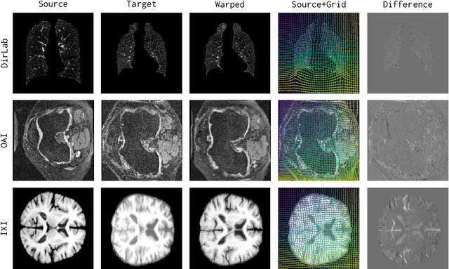
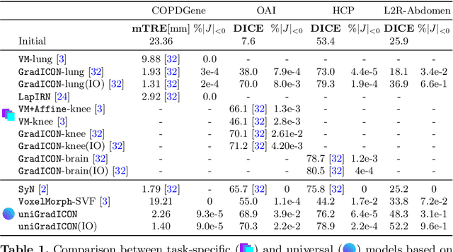
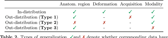
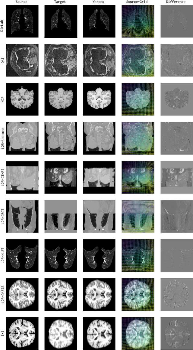
Conventional medical image registration approaches directly optimize over the parameters of a transformation model. These approaches have been highly successful and are used generically for registrations of different anatomical regions. Recent deep registration networks are incredibly fast and accurate but are only trained for specific tasks. Hence, they are no longer generic registration approaches. We therefore propose uniGradICON, a first step toward a foundation model for registration providing 1) great performance \emph{across} multiple datasets which is not feasible for current learning-based registration methods, 2) zero-shot capabilities for new registration tasks suitable for different acquisitions, anatomical regions, and modalities compared to the training dataset, and 3) a strong initialization for finetuning on out-of-distribution registration tasks. UniGradICON unifies the speed and accuracy benefits of learning-based registration algorithms with the generic applicability of conventional non-deep-learning approaches. We extensively trained and evaluated uniGradICON on twelve different public datasets. Our code and the uniGradICON model are available at https://github.com/uncbiag/uniGradICON.
Inverse Consistency by Construction for Multistep Deep Registration
Apr 28, 2023Inverse consistency is a desirable property for image registration. We propose a simple technique to make a neural registration network inverse consistent by construction, as a consequence of its structure, as long as it parameterizes its output transform by a Lie group. We extend this technique to multi-step neural registration by composing many such networks in a way that preserves inverse consistency. This multi-step approach also allows for inverse-consistent coarse to fine registration. We evaluate our technique on synthetic 2-D data and four 3-D medical image registration tasks and obtain excellent registration accuracy while assuring inverse consistency.