 Add to Chrome
 Add to Firefox
 Add to Edge
XRF-to-Optical Field-of-View Localization with Vision Language Models
Aug 18, 2026
Registering images acquired with different microscopy modalities is essential for relating complementary measurements of the same specimen. In correlative X-ray fluorescence (XRF) and optical microscopy, the XRF map often covers only a small region of an optical image acquired from the same or an adjacent tissue section. Field-of-view (FOV) localization is necessary but can be difficult when appearance and structure differ across modalities. Here we evaluate training-free vision language model (VLM) localization on two datasets representing same-section high-correspondence and adjacent-section low-correspondence imaging. We test unconstrained and metadata-constrained search and compare VLMs with geometric controls, classical template matching, and two alternative training-free approaches (DINOv2 and multiGradICON). Direct VLM prompting produced content-dependent spatial signals but was not reliable alone. Classical matching was most accurate when cross-modal structure was preserved but failed in the low-correspondence collection. A proposal-and-verify workflow used repeated VLM predictions as candidates and image-based similarity to select the final location. This workflow recovered useful localization in the low-correspondence regime.

Acquisition Geometry-Assisted Whole-Group Localization of X-ray Fluorescence Maps in Optical Microscopy Images
Aug 18, 2026X-ray fluorescence (XRF) microscopy maps elemental distributions, while optical microscopy can provide complementary morphological context. Localizing XRF fields of view (FOVs) in optical images is difficult because the two modalities differ in contrast mechanism and resolution. Most current workflows place each XRF tile independently, even when acquisition metadata already record the tiles' relative scan positions. This study formalizes XRF tile-group localization, in which one optical-frame placement is estimated for the whole group, constrained by acquisition geometry and quantified using group intersection-over-union (GroupIoU). In a controlled case study, independent localization failed with GroupIoU 0.000, whereas group localization achieved 0.931. Replacing the normalized cross-correlation (NCC) metric with mutual information (MI) gave nearly identical results, showing that the outcome is not specific to one local similarity metric. In another multiscale case study, using a coarse XRF survey scan to connect the fine-scale tile group to the optical image increased mean GroupIoU from 0.694 to 0.856. These case studies support using acquisition geometry as an explicit constraint when localizing related XRF tiles.
Accelerated deep self-supervised ptycho-laminography for three-dimensional nanoscale imaging of integrated circuits
Apr 10, 2023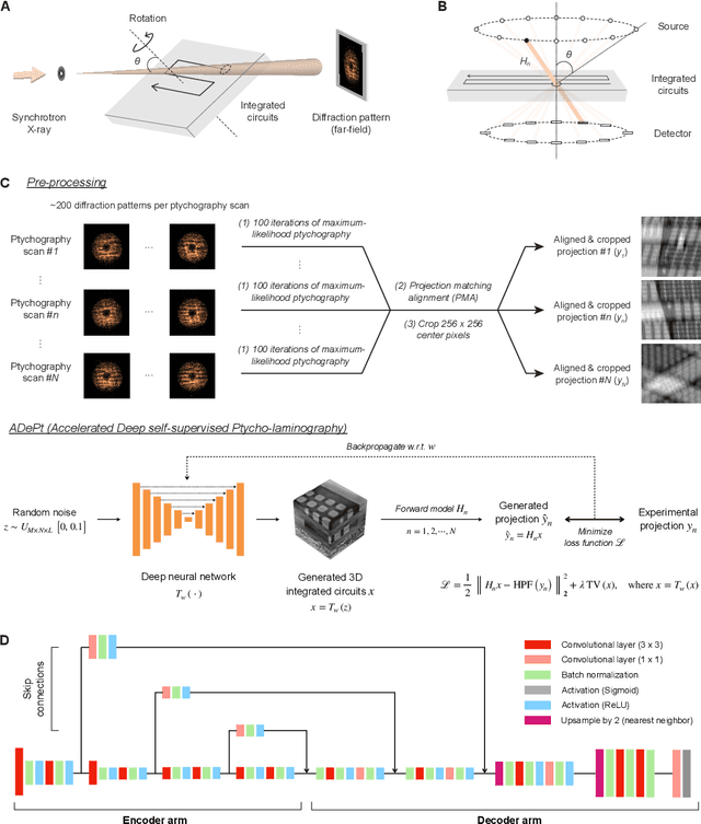
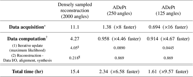
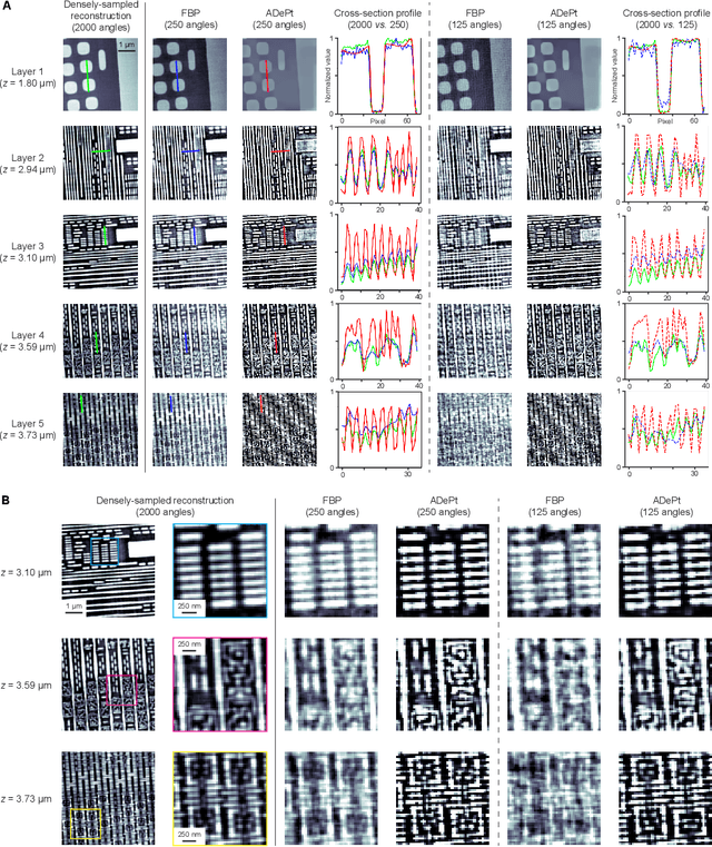
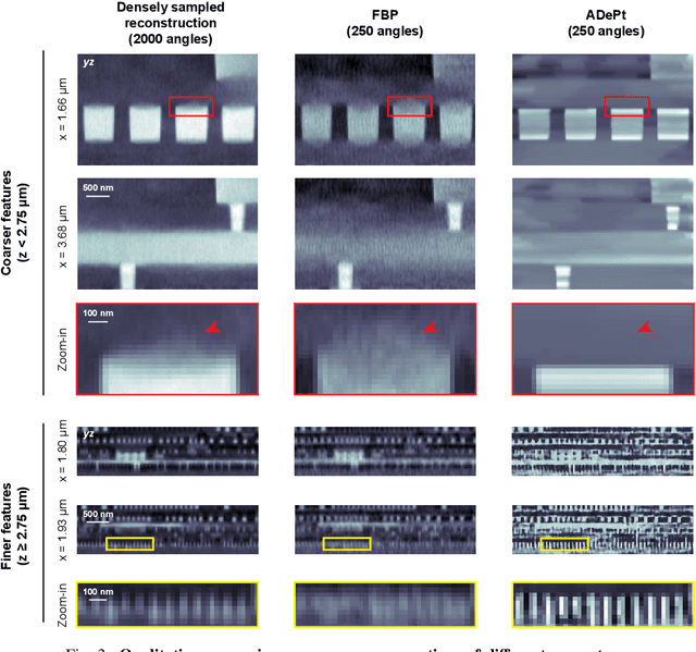
Three-dimensional inspection of nanostructures such as integrated circuits is important for security and reliability assurance. Two scanning operations are required: ptychographic to recover the complex transmissivity of the specimen; and rotation of the specimen to acquire multiple projections covering the 3D spatial frequency domain. Two types of rotational scanning are possible: tomographic and laminographic. For flat, extended samples, for which the full 180 degree coverage is not possible, the latter is preferable because it provides better coverage of the 3D spatial frequency domain compared to limited-angle tomography. It is also because the amount of attenuation through the sample is approximately the same for all projections. However, both techniques are time consuming because of extensive acquisition and computation time. Here, we demonstrate the acceleration of ptycho-laminographic reconstruction of integrated circuits with 16-times fewer angular samples and 4.67-times faster computation by using a physics-regularized deep self-supervised learning architecture. We check the fidelity of our reconstruction against a densely sampled reconstruction that uses full scanning and no learning. As already reported elsewhere [Zhou and Horstmeyer, Opt. Express, 28(9), pp. 12872-12896], we observe improvement of reconstruction quality even over the densely sampled reconstruction, due to the ability of the self-supervised learning kernel to fill the missing cone.
Attentional Ptycho-Tomography (APT) for three-dimensional nanoscale X-ray imaging with minimal data acquisition and computation time
Nov 29, 2022Noninvasive X-ray imaging of nanoscale three-dimensional objects, e.g. integrated circuits (ICs), generally requires two types of scanning: ptychographic, which is translational and returns estimates of complex electromagnetic field through ICs; and tomographic scanning, which collects complex field projections from multiple angles. Here, we present Attentional Ptycho-Tomography (APT), an approach trained to provide accurate reconstructions of ICs despite incomplete measurements, using a dramatically reduced amount of angular scanning. Training process includes regularizing priors based on typical IC patterns and the physics of X-ray propagation. We demonstrate that APT with 12-time reduced angles achieves fidelity comparable to the gold standard with the original set of angles. With the same set of reduced angles, APT also outperforms baseline reconstruction methods. In our experiments, APT achieves 108-time aggregate reduction in data acquisition and computation without compromising quality. We expect our physics-assisted machine learning framework could also be applied to other branches of nanoscale imaging.
Ptychopy: GPU framework for ptychographic data analysis
Jan 24, 2022X-ray ptychography imaging at synchrotron facilities like the Advanced Photon Source (APS) involves controlling instrument hardwares to collect a set of diffraction patterns from overlapping coherent illumination spots on extended samples, managing data storage, reconstructing ptychographic images from acquired diffraction patterns, and providing the visualization of results and feedback. In addition to the complicated workflow, ptychography instrument could produce up to several TB's of data per second that is needed to be processed in real time. This brings up the need to develop a high performance, robust and user friendly processing software package for ptychographic data analysis. In this paper we present a software framework which provides functionality of visualization, work flow control, and data reconstruction. To accelerate the computation and large datasets process, the data reconstruction part is implemented with three algorithms, ePIE, DM and LSQML using CUDA-C on GPU.