 Add to Chrome
 Add to Firefox
 Add to Edge
Rotational ultrasound and photoacoustic tomography of the human body
Apr 22, 2025Imaging the human body's morphological and angiographic information is essential for diagnosing, monitoring, and treating medical conditions. Ultrasonography performs the morphological assessment of the soft tissue based on acoustic impedance variations, whereas photoacoustic tomography (PAT) can visualize blood vessels based on intrinsic hemoglobin absorption. Three-dimensional (3D) panoramic imaging of the vasculature is generally not practical in conventional ultrasonography with limited field-of-view (FOV) probes, and PAT does not provide sufficient scattering-based soft tissue morphological contrast. Complementing each other, fast panoramic rotational ultrasound tomography (RUST) and PAT are integrated for hybrid rotational ultrasound and photoacoustic tomography (RUS-PAT), which obtains 3D ultrasound structural and PAT angiographic images of the human body quasi-simultaneously. The RUST functionality is achieved in a cost-effective manner using a single-element ultrasonic transducer for ultrasound transmission and rotating arc-shaped arrays for 3D panoramic detection. RUST is superior to conventional ultrasonography, which either has a limited FOV with a linear array or is high-cost with a hemispherical array that requires both transmission and receiving. By switching the acoustic source to a light source, the system is conveniently converted to PAT mode to acquire angiographic images in the same region. Using RUS-PAT, we have successfully imaged the human head, breast, hand, and foot with a 10 cm diameter FOV, submillimeter isotropic resolution, and 10 s imaging time for each modality. The 3D RUS-PAT is a powerful tool for high-speed, 3D, dual-contrast imaging of the human body with potential for rapid clinical translation.
Single-shot 3D photoacoustic computed tomography with a densely packed array for transcranial functional imaging
Jun 26, 2023


Photoacoustic computed tomography (PACT) is emerging as a new technique for functional brain imaging, primarily due to its capabilities in label-free hemodynamic imaging. Despite its potential, the transcranial application of PACT has encountered hurdles, such as acoustic attenuations and distortions by the skull and limited light penetration through the skull. To overcome these challenges, we have engineered a PACT system that features a densely packed hemispherical ultrasonic transducer array with 3072 channels, operating at a central frequency of 1 MHz. This system allows for single-shot 3D imaging at a rate equal to the laser repetition rate, such as 20 Hz. We have achieved a single-shot light penetration depth of approximately 9 cm in chicken breast tissue utilizing a 750 nm laser (withstanding 3295-fold light attenuation and still retaining an SNR of 74) and successfully performed transcranial imaging through an ex vivo human skull using a 1064 nm laser. Moreover, we have proven the capacity of our system to perform single-shot 3D PACT imaging in both tissue phantoms and human subjects. These results suggest that our PACT system is poised to unlock potential for real-time, in vivo transcranial functional imaging in humans.
Transcranial photoacoustic computed tomography of human brain function
Jun 01, 2022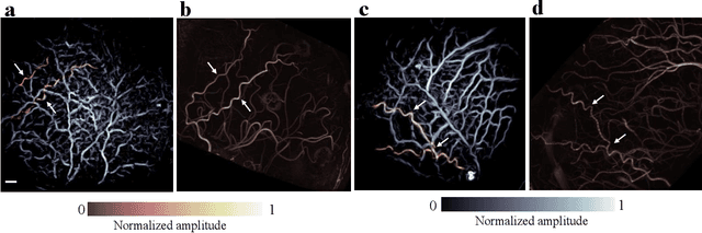
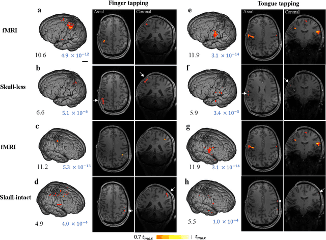
Herein we report the first in-human transcranial imaging of brain function using photoacoustic computed tomography. Functional responses to benchmark motor tasks were imaged on both the skull-less and the skull-intact hemispheres of a hemicraniectomy patient. The observed brain responses in these preliminary results demonstrate the potential of photoacoustic computed tomography for achieving transcranial functional imaging.