 Add to Chrome
 Add to Firefox
 Add to Edge
Harmonization in Magnetic Resonance Imaging: A Survey of Acquisition, Image-level, and Feature-level Methods
Jul 22, 2025Modern medical imaging technologies have greatly advanced neuroscience research and clinical diagnostics. However, imaging data collected across different scanners, acquisition protocols, or imaging sites often exhibit substantial heterogeneity, known as "batch effects" or "site effects". These non-biological sources of variability can obscure true biological signals, reduce reproducibility and statistical power, and severely impair the generalizability of learning-based models across datasets. Image harmonization aims to eliminate or mitigate such site-related biases while preserving meaningful biological information, thereby improving data comparability and consistency. This review provides a comprehensive overview of key concepts, methodological advances, publicly available datasets, current challenges, and future directions in the field of medical image harmonization, with a focus on magnetic resonance imaging (MRI). We systematically cover the full imaging pipeline, and categorize harmonization approaches into prospective acquisition and reconstruction strategies, retrospective image-level and feature-level methods, and traveling-subject-based techniques. Rather than providing an exhaustive survey, we focus on representative methods, with particular emphasis on deep learning-based approaches. Finally, we summarize the major challenges that remain and outline promising avenues for future research.
High-efficient deep learning-based DTI reconstruction with flexible diffusion gradient encoding scheme
Aug 02, 2023Purpose: To develop and evaluate a novel dynamic-convolution-based method called FlexDTI for high-efficient diffusion tensor reconstruction with flexible diffusion encoding gradient schemes. Methods: FlexDTI was developed to achieve high-quality DTI parametric mapping with flexible number and directions of diffusion encoding gradients. The proposed method used dynamic convolution kernels to embed diffusion gradient direction information into feature maps of the corresponding diffusion signal. Besides, our method realized the generalization of a flexible number of diffusion gradient directions by setting the maximum number of input channels of the network. The network was trained and tested using data sets from the Human Connectome Project and a local hospital. Results from FlexDTI and other advanced tensor parameter estimation methods were compared. Results: Compared to other methods, FlexDTI successfully achieves high-quality diffusion tensor-derived variables even if the number and directions of diffusion encoding gradients are variable. It increases peak signal-to-noise ratio (PSNR) by about 10 dB on Fractional Anisotropy (FA) and Mean Diffusivity (MD), compared with the state-of-the-art deep learning method with flexible diffusion encoding gradient schemes. Conclusion: FlexDTI can well learn diffusion gradient direction information to achieve generalized DTI reconstruction with flexible diffusion gradient schemes. Both flexibility and reconstruction quality can be taken into account in this network.
High-efficient Bloch simulation of magnetic resonance imaging sequences based on deep learning
Oct 19, 2022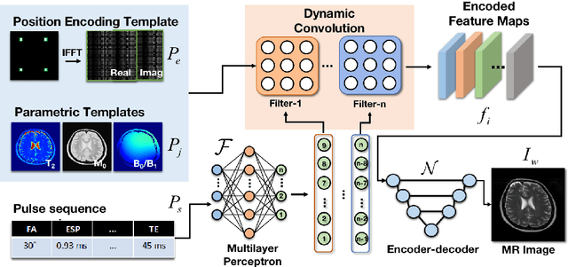
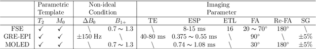
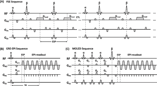
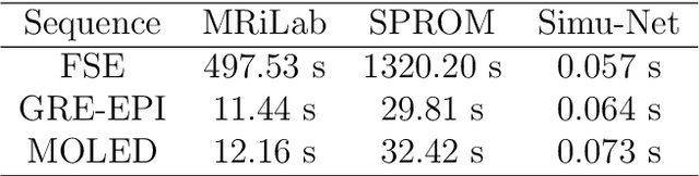
Bloch simulation constitutes an essential part of magnetic resonance imaging (MRI) development. However, even with the graphics processing units (GPU) acceleration, the heavy computational load remains a major challenge, especially in large-scale, high-accuracy simulation scenarios. Here we present a framework based on convolutional neural networks to build a high-efficient 2D Bloch simulator, termed Simu-Net. Compared to the mainstream GPU-based MRI simulation software, Simu-Net successfully accelerates simulations by over hundreds of times in three MRI pulse sequences. The accuracy and robustness of the proposed framework were also verified qualitatively and quantitatively. The trained Simu-Net was applied to generate sufficient customized training samples for deep learning-based T2 mapping and comparable results to conventional methods were obtained in the human brain. As a proof-of-concept work, Simu-Net shows the potential to apply deep learning for rapidly approximating the Bloch equation as a forward physical process.
Physics-driven Synthetic Data Learning for Biomedical Magnetic Resonance
Mar 22, 2022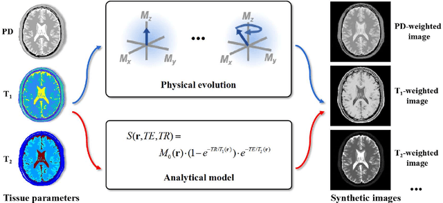
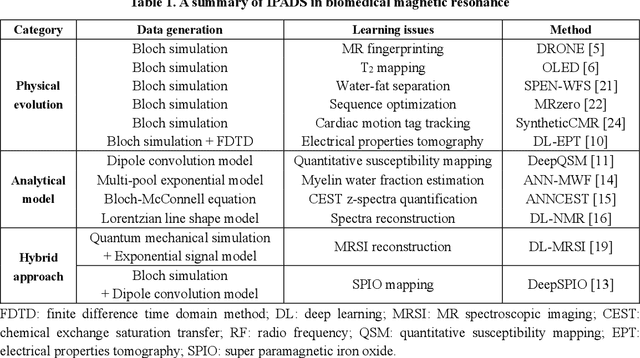
Deep learning has innovated the field of computational imaging. One of its bottlenecks is unavailable or insufficient training data. This article reviews an emerging paradigm, imaging physics-based data synthesis (IPADS), that can provide huge training data in biomedical magnetic resonance without or with few real data. Following the physical law of magnetic resonance, IPADS generates signals from differential equations or analytical solution models, making the learning more scalable, explainable, and better protecting privacy. Key components of IPADS learning, including signal generation models, basic deep learning network structures, enhanced data generation, and learning methods are discussed. Great potentials of IPADS have been demonstrated by representative applications in fast imaging, ultrafast signal reconstruction and accurate parameter quantification. Finally, open questions and future work have been discussed.
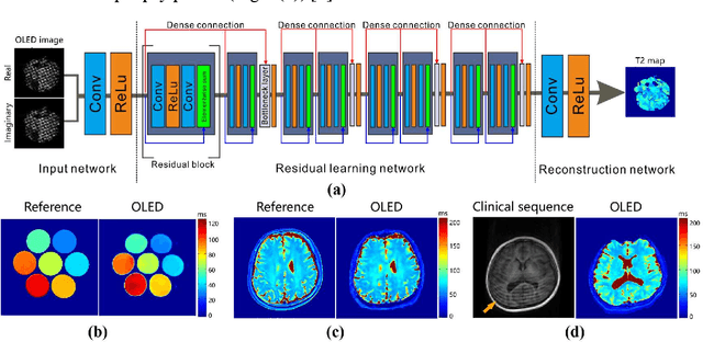
Model-based Synthetic Data-driven Learning (MOST-DL): Application in Single-shot T2 Mapping with Severe Head Motion Using Overlapping-echo Acquisition
Jul 30, 2021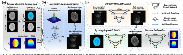
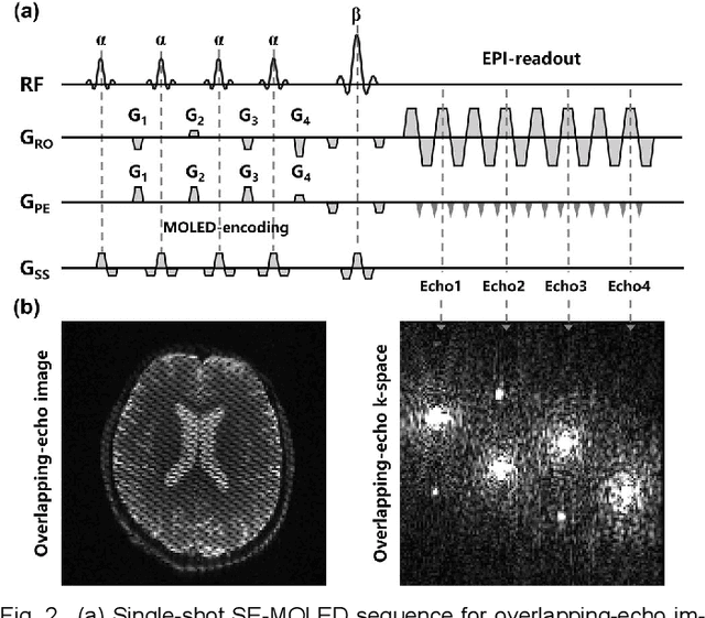
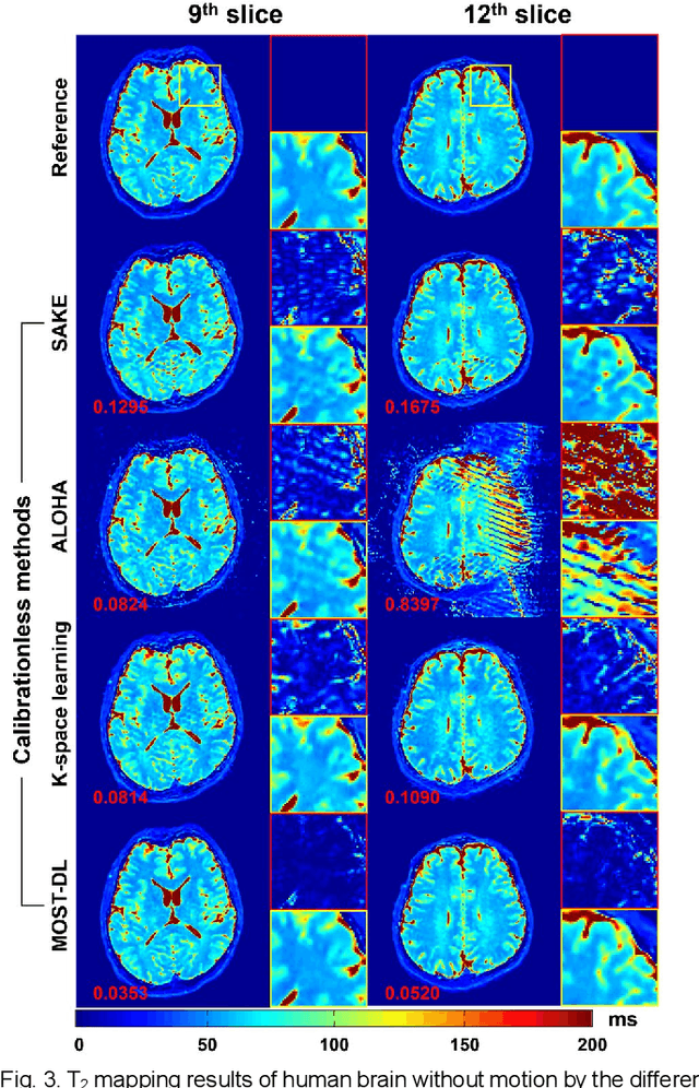
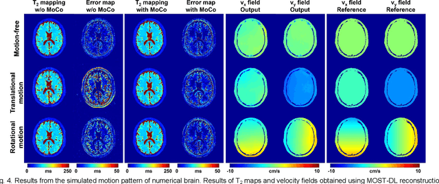
Data-driven learning algorithm has been successfully applied to facilitate reconstruction of medical imaging. However, real-world data needed for supervised learning are typically unavailable or insufficient, especially in the field of magnetic resonance imaging (MRI). Synthetic training samples have provided a potential solution for such problem, while the challenge brought by various non-ideal situations were usually encountered especially under complex experimental conditions. In this study, a general framework, Model-based Synthetic Data-driven Learning (MOST-DL), was proposed to generate paring data for network training to achieve robust T2 mapping using overlapping-echo acquisition under severe head motion accompanied with inhomogeneous RF field. We decomposed this challenging task into parallel reconstruction and motion correction according to a forward model. The neural network was first trained in pure synthetic dataset and then evaluated with in vivo human brain. Experiments showed that MOST-DL method significantly reduces ghosting and motion artifacts in T2 maps in the presence of random and continuous subject movement. We believe that the proposed approach may open a door for solving similar problems with other MRI acquisition methods and can be extended to other areas of medical imaging.