 Add to Chrome
 Add to Firefox
 Add to Edge
UPolarSQ: Polar Representation Learning for Optic Disc and Peripapillary Atrophy Segmentation and Quantification in Fundus Photographs
Aug 09, 2026
Myopia-induced posterior-pole remodeling is frequently accompanied by Optic Disc (OD) deformation and Peripapillary Atrophy (PPA), both of which provide clinically relevant structural biomarkers. In Cartesian fundus images, however, PPA often appears as an irregular and partially visible crescent adjacent to the OD, leading to fragmented segmentation and post-processing-dependent quantification. We propose UPolarSQ, a unified polar-domain framework for OD/PPA segmentation and biomarker quantification in myopic fundus images. UPolarSQ first maps an OD-centered region of interest into polar coordinates, where OD and PPA boundaries can be represented as radial profiles. It then employs UPolarSeg, a U-Net-based segmentation network enhanced with a Radial-Angular-Decoupled Module and boundary-aware auxiliary supervision to model anisotropic polar features and radial boundary transitions. Clinical biomarkers, including disc shape and PPA-width-related measurements, are deterministically extracted from the predicted polar masks, aligning segmentation and quantification within a shared geometric representation. Experiments on internal and external cohorts demonstrate that UPolarSQ improves OD/PPA segmentation and supports reliable polar-native biomarker estimation for myopic analysis.

SpecF2M: A Spectral-Aware Multi-task Network Estimating Axial Length and Refractive Error from Pediatric Fundus Photographs
Aug 07, 2026Spherical Equivalent Refraction (SER) and Axial Length (AL) are core indicators for pediatric myopia screening, yet their measurements require dedicated biometry and cycloplegic refraction. Fundus photography offers an accessible imaging modality, as myopia-related posterior-pole changes are visible in 45$^\circ$ fundus images. However, these cues are often low-contrast, spatially diffuse, and multi-scale. Moreover, AL, Sphere (SPH), and Cylinder (CYL) share partially overlapping but non-identical anatomical correlates. We propose SpecF2M, a spectral-aware multi-task network for estimating AL and SER components from pediatric fundus photographs. SpecF2M integrates a deterministic anatomy-guided enhancement module, a hybrid spatial--spectral backbone combining MixCNN and Hybrid Spectral Learning (HSL) blocks, and an expert-routing head for component-level estimation of AL, SPH, and CYL. On a pediatric cohort of 4,359 eligible child visits and 6,966 fundus images, SpecF2M outperforms controlled CNN/ViT baselines for AL and SPH estimation, achieving MAEs of 0.5347 mm and 0.7062 D, respectively. Component-level analysis further reveals asymmetric task coupling, where CYL exhibits weaker association with fundus-derived myopic patterns than AL/SPH. These results support fundus-based, screening-oriented estimation of pediatric myopia indicators, while external validation remains necessary before deployment.
Artificial Intelligence for Biomedical Video Generation
Nov 12, 2024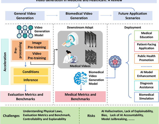
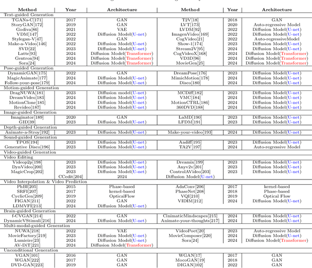
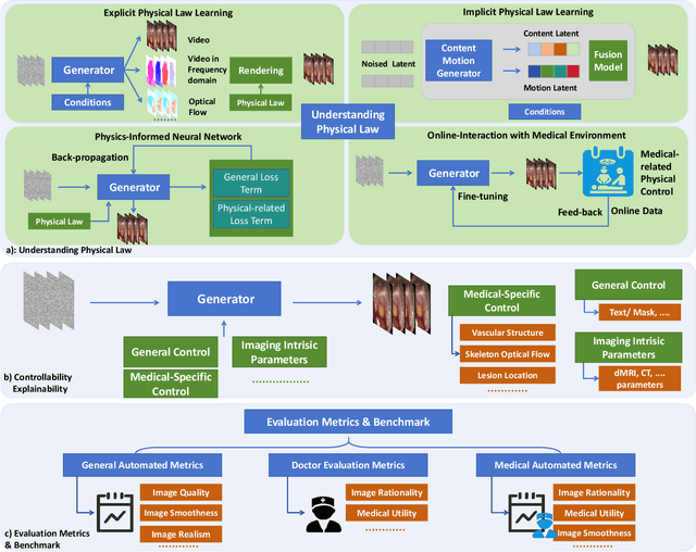
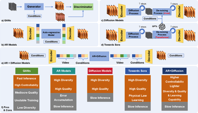
As a prominent subfield of Artificial Intelligence Generated Content (AIGC), video generation has achieved notable advancements in recent years. The introduction of Sora-alike models represents a pivotal breakthrough in video generation technologies, significantly enhancing the quality of synthesized videos. Particularly in the realm of biomedicine, video generation technology has shown immense potential such as medical concept explanation, disease simulation, and biomedical data augmentation. In this article, we thoroughly examine the latest developments in video generation models and explore their applications, challenges, and future opportunities in the biomedical sector. We have conducted an extensive review and compiled a comprehensive list of datasets from various sources to facilitate the development and evaluation of video generative models in biomedicine. Given the rapid progress in this field, we have also created a github repository to regularly update the advances of biomedical video generation at: https://github.com/Lee728243228/Biomedical-Video-Generation