 Add to Chrome
 Add to Firefox
 Add to Edge
Bridging MRI and PET physiology: Untangling complementarity through orthogonal representations
Apr 08, 2026
Multimodal imaging analysis often relies on joint latent representations, yet these approaches rarely define what information is shared versus modality-specific. Clarifying this distinction is clinically relevant, as it delineates the irreducible contribution of each modality and informs rational acquisition strategies. We propose a subspace decomposition framework that reframes multimodal fusion as a problem of orthogonal subspace separation rather than translation. We decompose Prostate-Specific Membrane Antigen (PSMA) PET uptake into an MRI-explainable physiological envelope and an orthogonal residual reflecting signal components not expressible within the MRI feature manifold. Using multiparametric MRI, we train an intensity-based, non-spatial implicit neural representation (INR) to map MRI feature vectors to PET uptake. We introduce a projection-based regularization using singular value decomposition to penalize residual components lying within the span of the MRI feature manifold. This enforces mathematical orthogonality between tissue-level physiological properties (structure, diffusion, perfusion) and intracellular PSMA expression. Tested on 13 prostate cancer patients, the model demonstrates that residual components spanned by MRI features are absorbed into the learned envelope, while the orthogonal residual is largest in tumour regions. This indicates that PSMA PET contains signal components not recoverable from MRI-derived physiological descriptors. The resulting decomposition provides a structured characterization of modality complementarity grounded in representation geometry rather than image translation.

Energy-based Tissue Manifolds for Longitudinal Multiparametric MRI Analysis
Apr 08, 2026We propose a geometric framework for longitudinal multi-parametric MRI analysis based on patient-specific energy modelling in sequence space. Rather than operating on images with spatial networks, each voxel is represented by its multi-sequence intensity vector ($T1$, $T1c$, $T2$, FLAIR, ADC), and a compact implicit neural representation is trained via denoising score matching to learn an energy function $E_θ(\mathbf{u})$ over $\mathbb{R}^d$ from a single baseline scan. The learned energy landscape provides a differential-geometric description of tissue regimes without segmentation labels. Local minima define tissue basins, gradient magnitude reflects proximity to regime boundaries, and Laplacian curvature characterises local constraint structure. Importantly, this baseline energy manifold is treated as a fixed geometric reference: it encodes the set of contrast combinations observed at diagnosis and is not retrained at follow-up. Longitudinal assessment is therefore formulated as evaluation of subsequent scans relative to this baseline geometry. Rather than comparing anatomical segmentations, we analyse how the distribution of MRI sequence vectors evolves under the baseline energy function. In a paediatric case with later recurrence, follow-up scans show progressive deviation in energy and directional displacement in sequence space toward the baseline tumour-associated regime before clear radiological reappearance. In a case with stable disease, voxel distributions remain confined to established low-energy basins without systematic drift. The presented cases serve as proof-of-concept that patient-specific energy manifolds can function as geometric reference systems for longitudinal mpMRI analysis without explicit segmentation or supervised classification, providing a foundation for further investigation of manifold-based tissue-at-risk tracking in neuro-oncology.
Beyond the LUMIR challenge: The pathway to foundational registration models
May 30, 2025
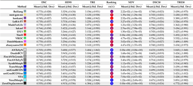
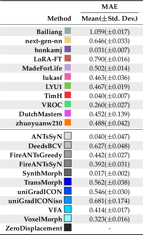
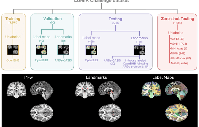
Medical image challenges have played a transformative role in advancing the field, catalyzing algorithmic innovation and establishing new performance standards across diverse clinical applications. Image registration, a foundational task in neuroimaging pipelines, has similarly benefited from the Learn2Reg initiative. Building on this foundation, we introduce the Large-scale Unsupervised Brain MRI Image Registration (LUMIR) challenge, a next-generation benchmark designed to assess and advance unsupervised brain MRI registration. Distinct from prior challenges that leveraged anatomical label maps for supervision, LUMIR removes this dependency by providing over 4,000 preprocessed T1-weighted brain MRIs for training without any label maps, encouraging biologically plausible deformation modeling through self-supervision. In addition to evaluating performance on 590 held-out test subjects, LUMIR introduces a rigorous suite of zero-shot generalization tasks, spanning out-of-domain imaging modalities (e.g., FLAIR, T2-weighted, T2*-weighted), disease populations (e.g., Alzheimer's disease), acquisition protocols (e.g., 9.4T MRI), and species (e.g., macaque brains). A total of 1,158 subjects and over 4,000 image pairs were included for evaluation. Performance was assessed using both segmentation-based metrics (Dice coefficient, 95th percentile Hausdorff distance) and landmark-based registration accuracy (target registration error). Across both in-domain and zero-shot tasks, deep learning-based methods consistently achieved state-of-the-art accuracy while producing anatomically plausible deformation fields. The top-performing deep learning-based models demonstrated diffeomorphic properties and inverse consistency, outperforming several leading optimization-based methods, and showing strong robustness to most domain shifts, the exception being a drop in performance on out-of-domain contrasts.
Fine-Tuning TransMorph with Gradient Correlation for Anatomical Alignment
Dec 30, 2024
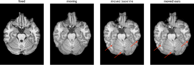
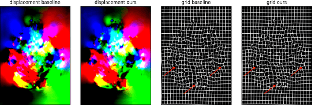
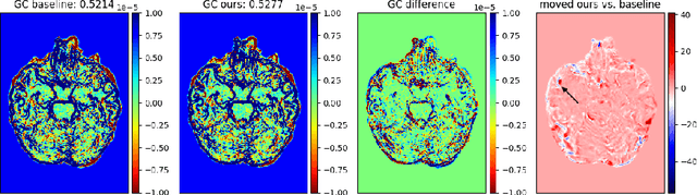
Unsupervised deep learning is a promising method in brain MRI registration to reduce the reliance on anatomical labels, while still achieving anatomically accurate transformations. For the Learn2Reg2024 LUMIR challenge, we propose fine-tuning of the pre-trained TransMorph model to improve the convergence stability as well as the deformation smoothness. The former is achieved through the FAdam optimizer, and consistency in structural changes is incorporated through the addition of gradient correlation in the similarity measure, improving anatomical alignment. The results show slight improvements in the Dice and HdDist95 scores, and a notable reduction in the NDV compared to the baseline TransMorph model. These are also confirmed by inspecting the boundaries of the tissue. Our proposed method highlights the effectiveness of including Gradient Correlation to achieve smoother and structurally consistent deformations for interpatient brain MRI registration.