 Add to Chrome
 Add to Firefox
 Add to Edge
MARVEL: Universal Murray's Law-informed Vessel Tree Segmentation and Topology Estimation
May 25, 2026
Vascular circulation follows fundamental biophysical principles that optimize mass transport and metabolic energy expenditure, which can be effectively modeled by Murray's law. However, contemporary deep learning methods for vascular segmentation often neglect these biophysical constraints. This leads to physiologically implausible branching and misclassification vascular trees, rendering. These automated segmentation results are unreliable unreliable for downstream clinical tasks such as blood flow simulation or disease quantification. In this paper, we introduce MARVEL (Universal MurrAy's law-infoRmed Vessel sEgmentation and topoLogy estimation), a backbone-agnostic framework that integrates biophysical priors into vascular tree extraction. MARVEL combines per-pixel supervision with explicit radius predictions to enforce local bifurcation constraints derived from an empirical width-exponent mapping. We implement these constraints as differentiable regularizers during training to guide models toward physiologically consistent reconstructions. We evaluate MARVEL on eight public datasets across multiple vascular modalities and segmentation backbones. Results demonstrate MARVEL's superior performance in segmentation accuracy, topological consistency, and physiological plausibility. By converting segmented masks into graph-based hemodynamic simulations, we demonstrate that MARVEL preserves the subtle pathological narrowing and topological connectivity required to distinguish hypertensive from normotensive eyes. Results show that MARVEL significantly improves the classification of hypertension via arteriovenous pressure differences in the eye (p < 0.001), outperforming baseline models in both topological consistency and clinical predictive value.

Are Macula or Optic Nerve Head Structures better at Diagnosing Glaucoma? An Answer using AI and Wide-Field Optical Coherence Tomography
Oct 13, 2022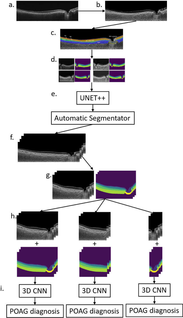
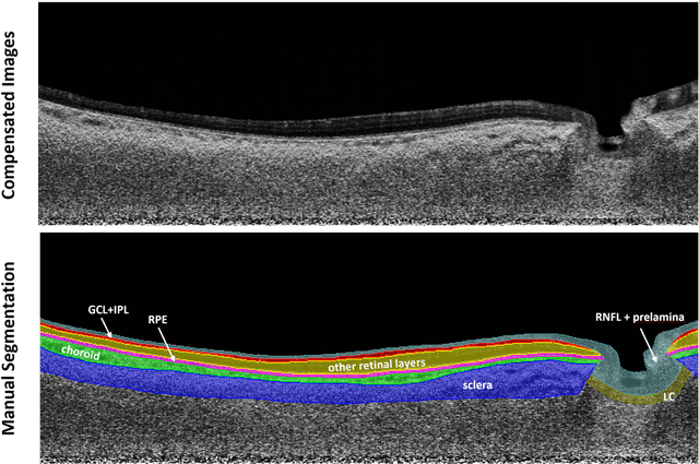
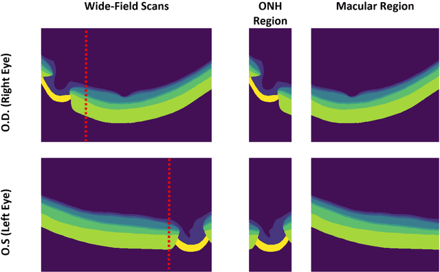
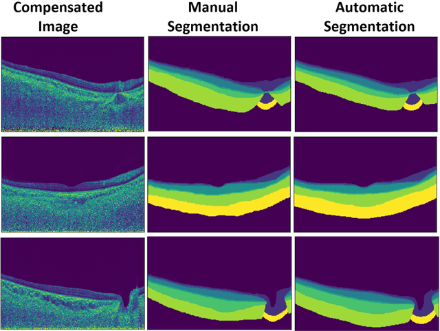
Purpose: (1) To develop a deep learning algorithm to automatically segment structures of the optic nerve head (ONH) and macula in 3D wide-field optical coherence tomography (OCT) scans; (2) To assess whether 3D macula or ONH structures (or the combination of both) provide the best diagnostic power for glaucoma. Methods: A cross-sectional comparative study was performed which included wide-field swept-source OCT scans from 319 glaucoma subjects and 298 non-glaucoma subjects. All scans were compensated to improve deep-tissue visibility. We developed a deep learning algorithm to automatically label all major ONH tissue structures by using 270 manually annotated B-scans for training. The performance of our algorithm was assessed using the Dice coefficient (DC). A glaucoma classification algorithm (3D CNN) was then designed using a combination of 500 OCT volumes and their corresponding automatically segmented masks. This algorithm was trained and tested on 3 datasets: OCT scans cropped to contain the macular tissues only, those to contain the ONH tissues only, and the full wide-field OCT scans. The classification performance for each dataset was reported using the AUC. Results: Our segmentation algorithm was able to segment ONH and macular tissues with a DC of 0.94 $\pm$ 0.003. The classification algorithm was best able to diagnose glaucoma using wide-field 3D-OCT volumes with an AUC of 0.99 $\pm$ 0.01, followed by ONH volumes with an AUC of 0.93 $\pm$ 0.06, and finally macular volumes with an AUC of 0.91 $\pm$ 0.11. Conclusions: this study showed that using wide-field OCT as compared to the typical OCT images containing just the ONH or macular may allow for a significantly improved glaucoma diagnosis. This may encourage the mainstream adoption of 3D wide-field OCT scans. For clinical AI studies that use traditional machines, we would recommend the use of ONH scans as opposed to macula scans.