 Add to Chrome
 Add to Firefox
 Add to Edge
Mitosis Detection in the Wild: Multi-Tumor and Context-Aware Generalization in the MIDOG 2025 Challenge
Jun 05, 2026Automated mitosis detection is a well-established task in computational pathology. While previous benchmarks focused on scanner-induced domain shift, clinical "real-world" application requires models to be robust across the vast variance to be expected in the histological landscape. The MItosis DOmain Generalization (MIDOG) 2025 challenge was designed to evaluate algorithmic performance across unprecedented biological and contextual diversity. We curated a test dataset of 365 cases, encompassing 12 distinct human, canine and feline tumor types, digitized across multiple scanning platforms. Moving beyond hand-selected hotspots, the challenge required detection also in random tissue areas (representative of the whole slide detection situation) and challenging areas (areas rich in hard negatives). In the second track, we introduced the classification of atypical mitotic figures (AMFs). There were 18 teams submitting to the detection track, with F1 scores ranging up to 0.740. In the AMF detection track, we had 21 submissions with balanced accuracy values up to 0.908. Our analysis reveals that while most models perform reliably in traditional hotspots, significant performance degradation occurs in challenging ROIs, where false positive rates tripled. Furthermore, performance varied significantly across the 12 tumor types, highlighting "blind spots" in current state-of-the-art architectures when encountering rare or highly pleomorphic malignancies. Moreover, we evaluated the effectiveness of ensembling and found a mean increases of 1.5 and 1.3 percentage points in F1 score and balanced accuracy, respectively. In contrast, TTA showed no relevant improvement. MIDOG 2025 demonstrates that "in the wild" mitosis detection remains a significant hurdle. The transition from hotspot-only evaluation to a multi-contextual framework provides a more realistic proxy for clinical reliability.
Z-Stack Scanning can Improve AI Detection of Mitosis: A Case Study of Meningiomas
Jan 27, 2025


Z-stack scanning is an emerging whole slide imaging technology that captures multiple focal planes alongside the z-axis of a glass slide. Because z-stacking can offer enhanced depth information compared to the single-layer whole slide imaging, this technology can be particularly useful in analyzing small-scaled histopathological patterns. However, its actual clinical impact remains debated with mixed results. To clarify this, we investigate the effect of z-stack scanning on artificial intelligence (AI) mitosis detection of meningiomas. With the same set of 22 Hematoxylin and Eosin meningioma glass slides scanned by three different digital pathology scanners, we tested the performance of three AI pipelines on both single-layer and z-stacked whole slide images (WSIs). Results showed that in all scanner-AI combinations, z-stacked WSIs significantly increased AI's sensitivity (+17.14%) on the mitosis detection with only a marginal impact on precision. Our findings provide quantitative evidence that highlights z-stack scanning as a promising technique for AI mitosis detection, paving the way for more reliable AI-assisted pathology workflows, which can ultimately benefit patient management.
Supporting Mitosis Detection AI Training with Inter-Observer Eye-Gaze Consistencies
Apr 02, 2024

The expansion of artificial intelligence (AI) in pathology tasks has intensified the demand for doctors' annotations in AI development. However, collecting high-quality annotations from doctors is costly and time-consuming, creating a bottleneck in AI progress. This study investigates eye-tracking as a cost-effective technology to collect doctors' behavioral data for AI training with a focus on the pathology task of mitosis detection. One major challenge in using eye-gaze data is the low signal-to-noise ratio, which hinders the extraction of meaningful information. We tackled this by levering the properties of inter-observer eye-gaze consistencies and creating eye-gaze labels from consistent eye-fixations shared by a group of observers. Our study involved 14 non-medical participants, from whom we collected eye-gaze data and generated eye-gaze labels based on varying group sizes. We assessed the efficacy of such eye-gaze labels by training Convolutional Neural Networks (CNNs) and comparing their performance to those trained with ground truth annotations and a heuristic-based baseline. Results indicated that CNNs trained with our eye-gaze labels closely followed the performance of ground-truth-based CNNs, and significantly outperformed the baseline. Although primarily focused on mitosis, we envision that insights from this study can be generalized to other medical imaging tasks.
Domain generalization across tumor types, laboratories, and species -- insights from the 2022 edition of the Mitosis Domain Generalization Challenge
Sep 27, 2023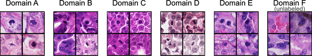
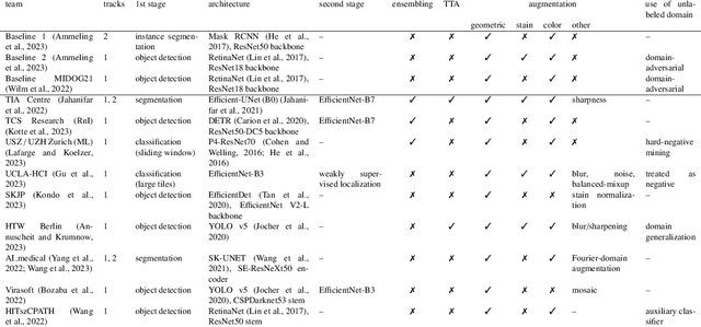
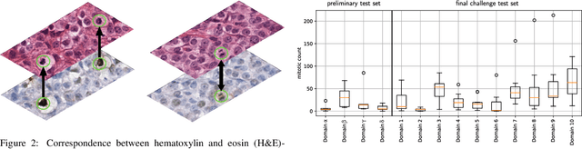
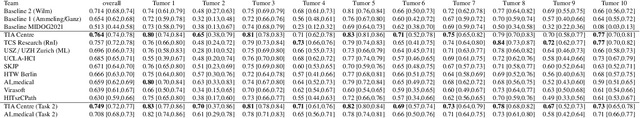
Recognition of mitotic figures in histologic tumor specimens is highly relevant to patient outcome assessment. This task is challenging for algorithms and human experts alike, with deterioration of algorithmic performance under shifts in image representations. Considerable covariate shifts occur when assessment is performed on different tumor types, images are acquired using different digitization devices, or specimens are produced in different laboratories. This observation motivated the inception of the 2022 challenge on MItosis Domain Generalization (MIDOG 2022). The challenge provided annotated histologic tumor images from six different domains and evaluated the algorithmic approaches for mitotic figure detection provided by nine challenge participants on ten independent domains. Ground truth for mitotic figure detection was established in two ways: a three-expert consensus and an independent, immunohistochemistry-assisted set of labels. This work represents an overview of the challenge tasks, the algorithmic strategies employed by the participants, and potential factors contributing to their success. With an $F_1$ score of 0.764 for the top-performing team, we summarize that domain generalization across various tumor domains is possible with today's deep learning-based recognition pipelines. When assessed against the immunohistochemistry-assisted reference standard, all methods resulted in reduced recall scores, but with only minor changes in the order of participants in the ranking.
Detecting Mitoses with a Convolutional Neural Network for MIDOG 2022 Challenge
Aug 26, 2022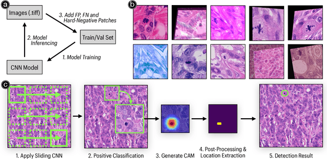
This work presents a mitosis detection method with only one vanilla Convolutional Neural Network (CNN). Our approach consists of two steps: given an image, we first apply a CNN using a sliding window technique to extract patches that have mitoses; we then calculate each extracted patch's class activation map to obtain the mitosis's precise location. To increase the model generalizability, we train the CNN with a series of data augmentation techniques, a loss that copes with noise-labeled images, and an active learning strategy. Our approach achieved an F1 score of 0.7323 with an EfficientNet-b3 model in the preliminary test phase of the MIDOG 2022 challenge.