 Add to Chrome
 Add to Firefox
 Add to Edge
Physical Embodiment Enables Information Processing Beyond Explicit Sensing in Active Matter
Aug 25, 2025Living microorganisms have evolved dedicated sensory machinery to detect environmental perturbations, processing these signals through biochemical networks to guide behavior. Replicating such capabilities in synthetic active matter remains a fundamental challenge. Here, we demonstrate that synthetic active particles can adapt to hidden hydrodynamic perturbations through physical embodiment alone, without explicit sensing mechanisms. Using reinforcement learning to control self-thermophoretic particles, we show that they learn navigation strategies to counteract unobserved flow fields by exploiting information encoded in their physical dynamics. Remarkably, particles successfully navigate perturbations that are not included in their state inputs, revealing that embodied dynamics can serve as an implicit sensing mechanism. This discovery establishes physical embodiment as a computational resource for information processing in active matter, with implications for autonomous microrobotic systems and bio-inspired computation.
Harnessing Synthetic Active Particles for Physical Reservoir Computing
Jul 27, 2023


The processing of information is an indispensable property of living systems realized by networks of active processes with enormous complexity. They have inspired many variants of modern machine learning one of them being reservoir computing, in which stimulating a network of nodes with fading memory enables computations and complex predictions. Reservoirs are implemented on computer hardware, but also on unconventional physical substrates such as mechanical oscillators, spins, or bacteria often summarized as physical reservoir computing. Here we demonstrate physical reservoir computing with a synthetic active microparticle system that self-organizes from an active and passive component into inherently noisy nonlinear dynamical units. The self-organization and dynamical response of the unit is the result of a delayed propulsion of the microswimmer to a passive target. A reservoir of such units with a self-coupling via the delayed response can perform predictive tasks despite the strong noise resulting from Brownian motion of the microswimmers. To achieve efficient noise suppression, we introduce a special architecture that uses historical reservoir states for output. Our results pave the way for the study of information processing in synthetic self-organized active particle systems.
Roadmap on Deep Learning for Microscopy
Mar 07, 2023


Through digital imaging, microscopy has evolved from primarily being a means for visual observation of life at the micro- and nano-scale, to a quantitative tool with ever-increasing resolution and throughput. Artificial intelligence, deep neural networks, and machine learning are all niche terms describing computational methods that have gained a pivotal role in microscopy-based research over the past decade. This Roadmap is written collectively by prominent researchers and encompasses selected aspects of how machine learning is applied to microscopy image data, with the aim of gaining scientific knowledge by improved image quality, automated detection, segmentation, classification and tracking of objects, and efficient merging of information from multiple imaging modalities. We aim to give the reader an overview of the key developments and an understanding of possibilities and limitations of machine learning for microscopy. It will be of interest to a wide cross-disciplinary audience in the physical sciences and life sciences.
Convolutional Neural Networks for Real-Time Localization and Classification in Feedback Digital Microscopy
Apr 10, 2020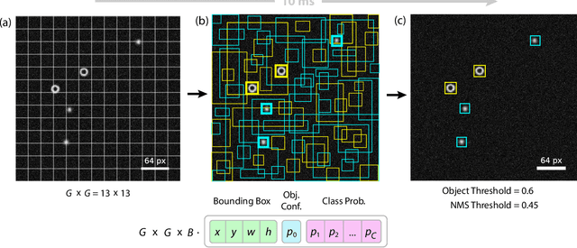
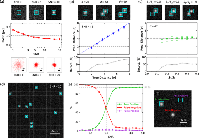
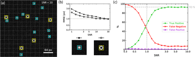
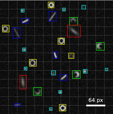
We present an adapted single-shot convolutional neural network (YOLOv2) for the real-time localization and classification of particles in optical microscopy. As compared to previous works, we focus on the real-time detection capabilities of the system to allow for manipulation of microscopic objects in large heterogeneous ensembles with the help of feedback control. The network is capable of localizing and classifying several hundreds of microscopic objects even at very low signal-to-noise ratios for images as large as 416x416 pixels with an inference time of about 10 ms. We demonstrate the real-time detection performance by manipulating active particles propelled by laser-induced self-thermophoresis. In order to make our framework readily available for others, we provide all scripts and source code. The network is implemented in Python/Keras using the TensorFlow backend. A C library supporting GPUs is provided for the real-time inference.