 Add to Chrome
 Add to Firefox
 Add to Edge
A geometry aware framework enhances noninvasive mapping of whole human brain dynamics
Apr 28, 2026
Non-invasive electrophysiology lacks methods that accurately reconstruct whole-brain spatiotemporal dynamics while incorporating individual cortical geometry, leaving current electroencephalography and magnetoencephalography source imaging limited by simplistic or biologically implausible priors. Here, we show that embedding participant-specific Geometric Basis Functions (GBFs), eigenmodes derived from each individual's cortical surface, provides a powerful anatomic constraint that resolves the inverse problem and improves reconstruction fidelity. The method reconstructs neural sources as linear combinations of geometric basis functions, thereby aligning source estimates with the geometric organization of neural dynamics. We validate GBF across the Meta-Source Benchmark, task-evoked data, resting-state networks, intracranial stimulation, and epilepsy data. The results demonstrate that GBF yields high localization accuracy and captures fast spatiotemporal dynamics consistent with anatomical pathways. These findings suggest that both spontaneous and evoked whole-brain activity can be described by hundreds of geometric modes, providing a compact yet accurate representation of neural sources. By linking cortical geometry to electrophysiological dynamics, GBF offers a versatile source imaging tool for both scientific and clinical applications.

Advancing EEG/MEG Source Imaging with Geometric-Informed Basis Functions
Jan 31, 2024

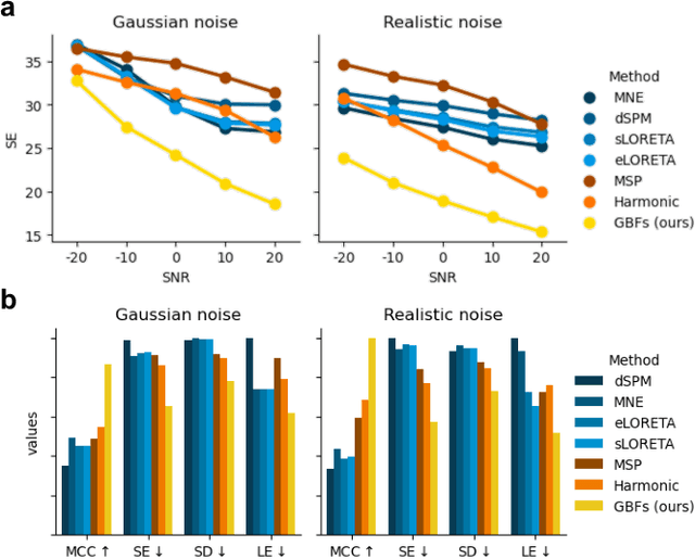

Electroencephalography (EEG) and Magnetoencephalography (MEG) are pivotal in understanding brain activity but are limited by their poor spatial resolution. EEG/MEG source imaging (ESI) infers the high-resolution electric field distribution in the brain based on the low-resolution scalp EEG/MEG observations. However, the ESI problem is ill-posed, and how to bring neuroscience priors into ESI method is the key. Here, we present a novel method which utilizes the Brain Geometric-informed Basis Functions (GBFs) as priors to enhance EEG/MEG source imaging. Through comprehensive experiments on both synthetic data and real task EEG data, we demonstrate the superiority of GBFs over traditional spatial basis functions (e.g., Harmonic and MSP), as well as existing ESI methods (e.g., dSPM, MNE, sLORETA, eLORETA). GBFs provide robust ESI results under different noise levels, and result in biologically interpretable EEG sources. We believe the high-resolution EEG source imaging from GBFs will greatly advance neuroscience research.