 Add to Chrome
 Add to Firefox
 Add to Edge
Revealing the Evolution of Order in Materials Microstructures Using Multi-Modal Computer Vision
Nov 15, 2024


The development of high-performance materials for microelectronics, energy storage, and extreme environments depends on our ability to describe and direct property-defining microstructural order. Our present understanding is typically derived from laborious manual analysis of imaging and spectroscopy data, which is difficult to scale, challenging to reproduce, and lacks the ability to reveal latent associations needed for mechanistic models. Here, we demonstrate a multi-modal machine learning (ML) approach to describe order from electron microscopy analysis of the complex oxide La$_{1-x}$Sr$_x$FeO$_3$. We construct a hybrid pipeline based on fully and semi-supervised classification, allowing us to evaluate both the characteristics of each data modality and the value each modality adds to the ensemble. We observe distinct differences in the performance of uni- and multi-modal models, from which we draw general lessons in describing crystal order using computer vision.
Final Report for CHESS: Cloud, High-Performance Computing, and Edge for Science and Security
Oct 21, 2024


Automating the theory-experiment cycle requires effective distributed workflows that utilize a computing continuum spanning lab instruments, edge sensors, computing resources at multiple facilities, data sets distributed across multiple information sources, and potentially cloud. Unfortunately, the obvious methods for constructing continuum platforms, orchestrating workflow tasks, and curating datasets over time fail to achieve scientific requirements for performance, energy, security, and reliability. Furthermore, achieving the best use of continuum resources depends upon the efficient composition and execution of workflow tasks, i.e., combinations of numerical solvers, data analytics, and machine learning. Pacific Northwest National Laboratory's LDRD "Cloud, High-Performance Computing (HPC), and Edge for Science and Security" (CHESS) has developed a set of interrelated capabilities for enabling distributed scientific workflows and curating datasets. This report describes the results and successes of CHESS from the perspective of open science.
SAM-I-Am: Semantic Boosting for Zero-shot Atomic-Scale Electron Micrograph Segmentation
Apr 09, 2024


Image segmentation is a critical enabler for tasks ranging from medical diagnostics to autonomous driving. However, the correct segmentation semantics - where are boundaries located? what segments are logically similar? - change depending on the domain, such that state-of-the-art foundation models can generate meaningless and incorrect results. Moreover, in certain domains, fine-tuning and retraining techniques are infeasible: obtaining labels is costly and time-consuming; domain images (micrographs) can be exponentially diverse; and data sharing (for third-party retraining) is restricted. To enable rapid adaptation of the best segmentation technology, we propose the concept of semantic boosting: given a zero-shot foundation model, guide its segmentation and adjust results to match domain expectations. We apply semantic boosting to the Segment Anything Model (SAM) to obtain microstructure segmentation for transmission electron microscopy. Our booster, SAM-I-Am, extracts geometric and textural features of various intermediate masks to perform mask removal and mask merging operations. We demonstrate a zero-shot performance increase of (absolute) +21.35%, +12.6%, +5.27% in mean IoU, and a -9.91%, -18.42%, -4.06% drop in mean false positive masks across images of three difficulty classes over vanilla SAM (ViT-L).
Deep Learning for Automated Experimentation in Scanning Transmission Electron Microscopy
Apr 04, 2023


Machine learning (ML) has become critical for post-acquisition data analysis in (scanning) transmission electron microscopy, (S)TEM, imaging and spectroscopy. An emerging trend is the transition to real-time analysis and closed-loop microscope operation. The effective use of ML in electron microscopy now requires the development of strategies for microscopy-centered experiment workflow design and optimization. Here, we discuss the associated challenges with the transition to active ML, including sequential data analysis and out-of-distribution drift effects, the requirements for the edge operation, local and cloud data storage, and theory in the loop operations. Specifically, we discuss the relative contributions of human scientists and ML agents in the ideation, orchestration, and execution of experimental workflows and the need to develop universal hyper languages that can apply across multiple platforms. These considerations will collectively inform the operationalization of ML in next-generation experimentation.
Design of a Graphical User Interface for Few-Shot Machine Learning Classification of Electron Microscopy Data
Jul 21, 2021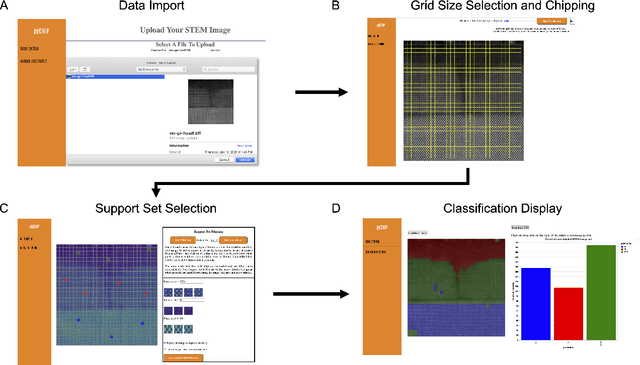
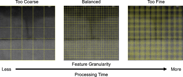

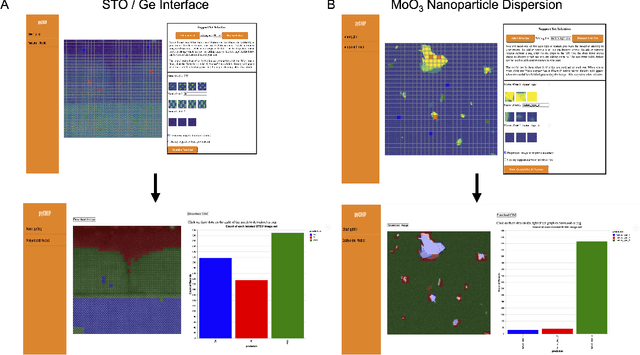
The recent growth in data volumes produced by modern electron microscopes requires rapid, scalable, and flexible approaches to image segmentation and analysis. Few-shot machine learning, which can richly classify images from a handful of user-provided examples, is a promising route to high-throughput analysis. However, current command-line implementations of such approaches can be slow and unintuitive to use, lacking the real-time feedback necessary to perform effective classification. Here we report on the development of a Python-based graphical user interface that enables end users to easily conduct and visualize the output of few-shot learning models. This interface is lightweight and can be hosted locally or on the web, providing the opportunity to reproducibly conduct, share, and crowd-source few-shot analyses.