 Add to Chrome
 Add to Firefox
 Add to Edge
X-LMC: Cross-View Spatiotemporal Collateral Circulation Scoring from DSA
Aug 19, 2026
Digital subtraction angiography (DSA) is the reference standard for leptomeningeal collateral (LMC) assessment, providing critical prognostic insights to guide secondary treatment strategies, neurorehabilitation planning, and retrospective stroke research. However, clinical LMC grading via the ASITN/SIR scale relies on manual, highly variable visual inspection. We introduce X-LMC, a spatiotemporal framework for automated collateral scoring from time-resolved biplane DSA. The proposed architecture encodes spatial frame representations through a DINOv2 backbone, fuses orthogonal projections via a token-level cross-view attention module, and models representations of contrast bolus dynamics using a recurrent network architecture. We evaluate our framework on a multicenter dataset of 134 patients with M1-segment occlusions. In a 5-fold cross-validation setting, X-LMC yields higher point estimates than static architectures and spatiotemporal baselines adapted from related angiographic tasks, achieving a Quadratic Weighted Kappa (QWK) of 0.398 (vs. 0.322) and a dichotomized macro-F1 score of 0.711 (vs. 0.663) against the best-performing baseline. X-LMC performance also aligns with the observed clinical inter-rater agreement (QWK: 0.314). As the first DSA study attempting to automate LMC scoring, we demonstrate that multi-view temporal deep learning can capture collateral-specific contrast kinetics. Ultimately, these benchmarks delineate the clinical ambiguities and achievable performance boundaries of automated ASITN/SIR grading, establishing a reproducible foundation for objective hemodynamic phenotyping in stroke cohorts. Code is available at https://github.com/maedehafezi/X-LMC.

Optimizing Rank for High-Fidelity Implicit Neural Representations
Dec 16, 2025Implicit Neural Representations (INRs) based on vanilla Multi-Layer Perceptrons (MLPs) are widely believed to be incapable of representing high-frequency content. This has directed research efforts towards architectural interventions, such as coordinate embeddings or specialized activation functions, to represent high-frequency signals. In this paper, we challenge the notion that the low-frequency bias of vanilla MLPs is an intrinsic, architectural limitation to learn high-frequency content, but instead a symptom of stable rank degradation during training. We empirically demonstrate that regulating the network's rank during training substantially improves the fidelity of the learned signal, rendering even simple MLP architectures expressive. Extensive experiments show that using optimizers like Muon, with high-rank, near-orthogonal updates, consistently enhances INR architectures even beyond simple ReLU MLPs. These substantial improvements hold across a diverse range of domains, including natural and medical images, and novel view synthesis, with up to 9 dB PSNR improvements over the previous state-of-the-art. Our project page, which includes code and experimental results, is available at: (https://muon-inrs.github.io).
Spatial Brain Tumor Concentration Estimation for Individualized Radiotherapy Planning
Dec 18, 2024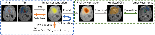
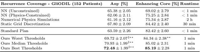
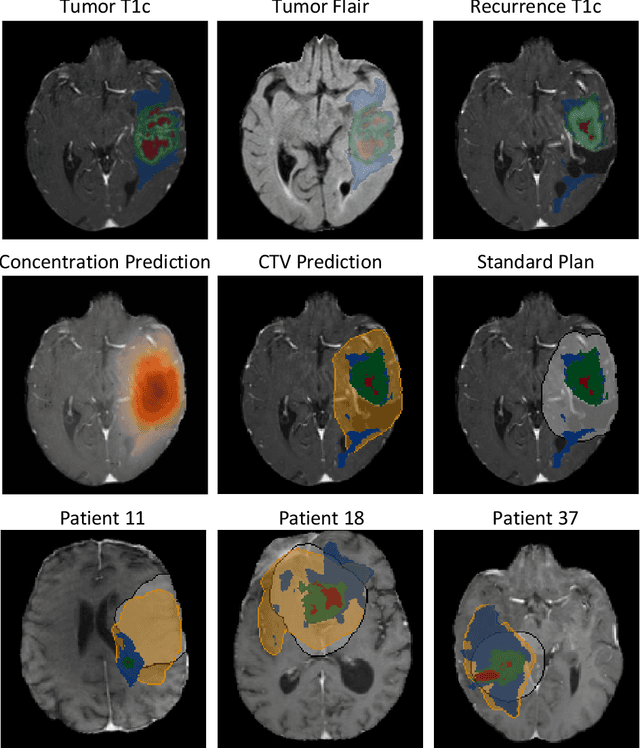
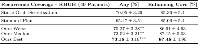
Biophysical modeling of brain tumors has emerged as a promising strategy for personalizing radiotherapy planning by estimating the otherwise hidden distribution of tumor cells within the brain. However, many existing state-of-the-art methods are computationally intensive, limiting their widespread translation into clinical practice. In this work, we propose an efficient and direct method that utilizes soft physical constraints to estimate the tumor cell concentration from preoperative MRI of brain tumor patients. Our approach optimizes a 3D tumor concentration field by simultaneously minimizing the difference between the observed MRI and a physically informed loss function. Compared to existing state-of-the-art techniques, our method significantly improves predicting tumor recurrence on two public datasets with a total of 192 patients while maintaining a clinically viable runtime of under one minute - a substantial reduction from the 30 minutes required by the current best approach. Furthermore, we showcase the generalizability of our framework by incorporating additional imaging information and physical constraints, highlighting its potential to translate to various medical diffusion phenomena with imperfect data.