 Add to Chrome
 Add to Firefox
 Add to Edge
Combining Cartesian and non-Cartesian acceleration techniques with SPARKLING for 1mm isotropic whole-brain MPRAGE in a minute
May 29, 2026Purpose: T1-weighted MPRAGE remains a cornerstone of clinical anatomical imaging, yet its long acquisition times constrain routine use. Established acceleration techniques, namely Parallel Imaging (PI) and Compressed Sensing (CS), tend to introduce substantial noise and blurring when pushed to high acceleration factors. Although they rely on fundamentally different redundancies, combining them synergistically remains an open challenge. Methods: The GoLF-SPARKLING framework was extended to jointly exploit two acceleration mechanisms: GRAPPA-based PI in the central k-space region and variable-density CS in the periphery, with independent acceleration factors in each zone. To preserve smooth signal evolution throughout the inversion-recovery period and avoid modulation artifacts, the acquisition trajectory was reordered accordingly. The resulting method was evaluated prospectively in vivo at 1mm isotropic resolution and benchmarked against Wave-CAIPI and Poisson-disk sampling. Results: The proposed hybrid approach produced sharper, less noisy, and more stable whole-brain images in approximately one minute than either acceleration strategy alone. Purely PI-based reconstructions were degraded by high g-factor noise, while purely CS-based reconstructions exhibited pronounced blurring. Furthermore, this method yielded lower average volumetric errors in downstream automated brain segmentation than state-of-the-art acceleration techniques, demonstrating its clinical utility. Conclusion: By jointly leveraging PI and CS, GoLF-SPARKLING achieves high acceleration factors that enable sub-minute, high-quality anatomical MRI. This translates into greater clinical throughput and more reliable imaging in patients who are challenging to scan.
SNAKE-fMRI: A modular fMRI data simulator from the space-time domain to k-space and back
Apr 12, 2024We propose a new, modular, open-source, Python-based 3D+time fMRI data simulation software, \emph{SNAKE-fMRI}, which stands for \emph{S}imulator from \emph{N}eurovascular coupling to \emph{A}cquisition of \emph{K}-space data for \emph{E}xploration of fMRI acquisition techniques.Unlike existing tools, the goal here is to simulate the complete chain of fMRI data acquisition, from the spatio-temporal design of evoked brain responses to various multi-coil k-space data 3D sampling strategies, with the possibility of extending the forward acquisition model to various noise and artifact sources while remaining memory-efficient.By using this \emph{in silico} setup, we are thus able to provide realistic and reproducible ground truth for fMRI reconstruction methods in 3D accelerated acquisition settings and explore the influence of critical parameters, such as the acceleration factor and signal-to-noise ratio~(SNR), on downstream tasks of image reconstruction and statistical analysis of evoked brain activity.We present three scenarios of increasing complexity to showcase the flexibility, versatility, and fidelity of \emph{SNAKE-fMRI}: From a temporally-fixed full 3D Cartesian to various 3D non-Cartesian sampling patterns, we can compare -- with reproducibility guarantees -- how experimental paradigms, acquisition strategies and reconstruction methods contribute and interact together, affecting the downstream statistical analysis.
Is good old GRAPPA dead?
Jun 01, 2021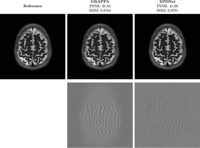
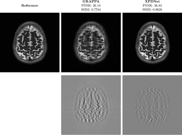
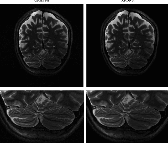
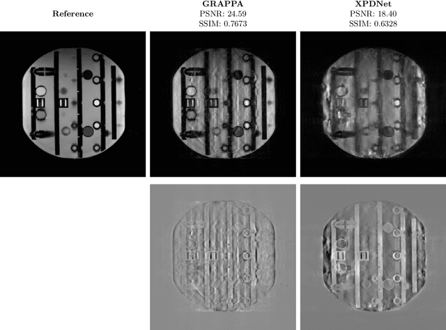
We perform a qualitative analysis of performance of XPDNet, a state-of-the-art deep learning approach for MRI reconstruction, compared to GRAPPA, a classical approach. We do this in multiple settings, in particular testing the robustness of the XPDNet to unseen settings, and show that the XPDNet can to some degree generalize well.
Robust imaging of hippocampal inner structure at 7T: in vivo acquisition protocol and methodological choices
May 09, 2016

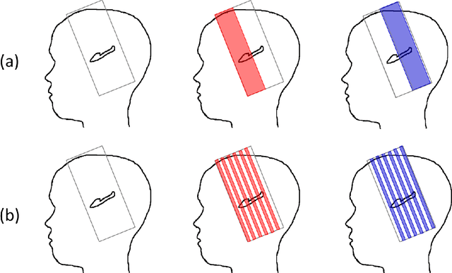
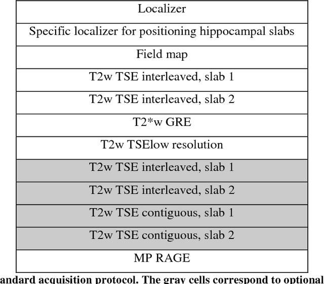
OBJECTIVE:Motion-robust multi-slab imaging of hippocampal inner structure in vivo at 7T.MATERIALS AND METHODS:Motion is a crucial issue for ultra-high resolution imaging, such as can be achieved with 7T MRI. An acquisition protocol was designed for imaging hippocampal inner structure at 7T. It relies on a compromise between anatomical details visibility and robustness to motion. In order to reduce acquisition time and motion artifacts, the full slab covering the hippocampus was split into separate slabs with lower acquisition time. A robust registration approach was implemented to combine the acquired slabs within a final 3D-consistent high-resolution slab covering the whole hippocampus. Evaluation was performed on 50 subjects overall, made of three groups of subjects acquired using three acquisition settings; it focused on three issues: visibility of hippocampal inner structure, robustness to motion artifacts and registration procedure performance.RESULTS:Overall, T2-weighted acquisitions with interleaved slabs proved robust. Multi-slab registration yielded high quality datasets in 96 % of the subjects, thus compatible with further analyses of hippocampal inner structure.CONCLUSION:Multi-slab acquisition and registration setting is efficient for reducing acquisition time and consequently motion artifacts for ultra-high resolution imaging of the inner structure of the hippocampus.