 Add to Chrome
 Add to Firefox
 Add to Edge
Mammogram
Papers and Code
Longitudinal Mammogram Risk Prediction
Apr 29, 2024


Breast cancer is one of the leading causes of mortality among women worldwide. Early detection and risk assessment play a crucial role in improving survival rates. Therefore, annual or biennial mammograms are often recommended for screening in high-risk groups. Mammograms are typically interpreted by expert radiologists based on the Breast Imaging Reporting and Data System (BI-RADS), which provides a uniform way to describe findings and categorizes them to indicate the level of concern for breast cancer. Recently, machine learning (ML) and computational approaches have been developed to automate and improve the interpretation of mammograms. However, both BI-RADS and the ML-based methods focus on the analysis of data from the present and sometimes the most recent prior visit. While it is clear that temporal changes in image features of the longitudinal scans should carry value for quantifying breast cancer risk, no prior work has conducted a systematic study of this. In this paper, we extend a state-of-the-art ML model to ingest an arbitrary number of longitudinal mammograms and predict future breast cancer risk. On a large-scale dataset, we demonstrate that our model, LoMaR, achieves state-of-the-art performance when presented with only the present mammogram. Furthermore, we use LoMaR to characterize the predictive value of prior visits. Our results show that longer histories (e.g., up to four prior annual mammograms) can significantly boost the accuracy of predicting future breast cancer risk, particularly beyond the short-term. Our code and model weights are available at https://github.com/batuhankmkaraman/LoMaR.
Mammo-CLIP: Leveraging Contrastive Language-Image Pre-training (CLIP) for Enhanced Breast Cancer Diagnosis with Multi-view Mammography
Apr 24, 2024Although fusion of information from multiple views of mammograms plays an important role to increase accuracy of breast cancer detection, developing multi-view mammograms-based computer-aided diagnosis (CAD) schemes still faces challenges and no such CAD schemes have been used in clinical practice. To overcome the challenges, we investigate a new approach based on Contrastive Language-Image Pre-training (CLIP), which has sparked interest across various medical imaging tasks. By solving the challenges in (1) effectively adapting the single-view CLIP for multi-view feature fusion and (2) efficiently fine-tuning this parameter-dense model with limited samples and computational resources, we introduce Mammo-CLIP, the first multi-modal framework to process multi-view mammograms and corresponding simple texts. Mammo-CLIP uses an early feature fusion strategy to learn multi-view relationships in four mammograms acquired from the CC and MLO views of the left and right breasts. To enhance learning efficiency, plug-and-play adapters are added into CLIP image and text encoders for fine-tuning parameters and limiting updates to about 1% of the parameters. For framework evaluation, we assembled two datasets retrospectively. The first dataset, comprising 470 malignant and 479 benign cases, was used for few-shot fine-tuning and internal evaluation of the proposed Mammo-CLIP via 5-fold cross-validation. The second dataset, including 60 malignant and 294 benign cases, was used to test generalizability of Mammo-CLIP. Study results show that Mammo-CLIP outperforms the state-of-art cross-view transformer in AUC (0.841 vs. 0.817, 0.837 vs. 0.807) on both datasets. It also surpasses previous two CLIP-based methods by 20.3% and 14.3%. This study highlights the potential of applying the finetuned vision-language models for developing next-generation, image-text-based CAD schemes of breast cancer.
Semi- and Weakly-Supervised Learning for Mammogram Mass Segmentation with Limited Annotations
Mar 14, 2024


Accurate identification of breast masses is crucial in diagnosing breast cancer; however, it can be challenging due to their small size and being camouflaged in surrounding normal glands. Worse still, it is also expensive in clinical practice to obtain adequate pixel-wise annotations for training deep neural networks. To overcome these two difficulties with one stone, we propose a semi- and weakly-supervised learning framework for mass segmentation that utilizes limited strongly-labeled samples and sufficient weakly-labeled samples to achieve satisfactory performance. The framework consists of an auxiliary branch to exclude lesion-irrelevant background areas, a segmentation branch for final prediction, and a spatial prompting module to integrate the complementary information of the two branches. We further disentangle encoded obscure features into lesion-related and others to boost performance. Experiments on CBIS-DDSM and INbreast datasets demonstrate the effectiveness of our method.
MV-Swin-T: Mammogram Classification with Multi-view Swin Transformer
Feb 26, 2024Traditional deep learning approaches for breast cancer classification has predominantly concentrated on single-view analysis. In clinical practice, however, radiologists concurrently examine all views within a mammography exam, leveraging the inherent correlations in these views to effectively detect tumors. Acknowledging the significance of multi-view analysis, some studies have introduced methods that independently process mammogram views, either through distinct convolutional branches or simple fusion strategies, inadvertently leading to a loss of crucial inter-view correlations. In this paper, we propose an innovative multi-view network exclusively based on transformers to address challenges in mammographic image classification. Our approach introduces a novel shifted window-based dynamic attention block, facilitating the effective integration of multi-view information and promoting the coherent transfer of this information between views at the spatial feature map level. Furthermore, we conduct a comprehensive comparative analysis of the performance and effectiveness of transformer-based models under diverse settings, employing the CBIS-DDSM and Vin-Dr Mammo datasets. Our code is publicly available at https://github.com/prithuls/MV-Swin-T
Adversarially Robust Feature Learning for Breast Cancer Diagnosis
Feb 13, 2024


Adversarial data can lead to malfunction of deep learning applications. It is essential to develop deep learning models that are robust to adversarial data while accurate on standard, clean data. In this study, we proposed a novel adversarially robust feature learning (ARFL) method for a real-world application of breast cancer diagnosis. ARFL facilitates adversarial training using both standard data and adversarial data, where a feature correlation measure is incorporated as an objective function to encourage learning of robust features and restrain spurious features. To show the effects of ARFL in breast cancer diagnosis, we built and evaluated diagnosis models using two independent clinically collected breast imaging datasets, comprising a total of 9,548 mammogram images. We performed extensive experiments showing that our method outperformed several state-of-the-art methods and that our method can enhance safer breast cancer diagnosis against adversarial attacks in clinical settings.
Learning using privileged information for segmenting tumors on digital mammograms
Feb 09, 2024


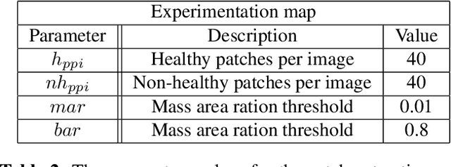
Limited amount of data and data sharing restrictions, due to GDPR compliance, constitute two common factors leading to reduced availability and accessibility when referring to medical data. To tackle these issues, we introduce the technique of Learning Using Privileged Information. Aiming to substantiate the idea, we attempt to build a robust model that improves the segmentation quality of tumors on digital mammograms, by gaining privileged information knowledge during the training procedure. Towards this direction, a baseline model, called student, is trained on patches extracted from the original mammograms, while an auxiliary model with the same architecture, called teacher, is trained on the corresponding enhanced patches accessing, in this way, privileged information. We repeat the student training procedure by providing the assistance of the teacher model this time. According to the experimental results, it seems that the proposed methodology performs better in the most of the cases and it can achieve 10% higher F1 score in comparison with the baseline.
AmbientCycleGAN for Establishing Interpretable Stochastic Object Models Based on Mathematical Phantoms and Medical Imaging Measurements
Feb 02, 2024Medical imaging systems that are designed for producing diagnostically informative images should be objectively assessed via task-based measures of image quality (IQ). Ideally, computation of task-based measures of IQ needs to account for all sources of randomness in the measurement data, including the variability in the ensemble of objects to be imaged. To address this need, stochastic object models (SOMs) that can generate an ensemble of synthesized objects or phantoms can be employed. Various mathematical SOMs or phantoms were developed that can interpretably synthesize objects, such as lumpy object models and parameterized torso phantoms. However, such SOMs that are purely mathematically defined may not be able to comprehensively capture realistic object variations. To establish realistic SOMs, it is desirable to use experimental data. An augmented generative adversarial network (GAN), AmbientGAN, was recently proposed for establishing SOMs from medical imaging measurements. However, it remains unclear to which extent the AmbientGAN-produced objects can be interpretably controlled. This work introduces a novel approach called AmbientCycleGAN that translates mathematical SOMs to realistic SOMs by use of noisy measurement data. Numerical studies that consider clustered lumpy background (CLB) models and real mammograms are conducted. It is demonstrated that our proposed method can stably establish SOMs based on mathematical models and noisy measurement data. Moreover, the ability of the proposed AmbientCycleGAN to interpretably control image features in the synthesized objects is investigated.
Siamese Networks with Soft Labels for Unsupervised Lesion Detection and Patch Pretraining on Screening Mammograms
Jan 10, 2024Self-supervised learning has become a popular way to pretrain a deep learning model and then transfer it to perform downstream tasks. However, most of these methods are developed on large-scale image datasets that contain natural objects with clear textures, outlines, and distinct color contrasts. It remains uncertain whether these methods are equally effective for medical imaging, where the regions of interest often blend subtly and indistinctly with the surrounding tissues. In this study, we propose an alternative method that uses contralateral mammograms to train a neural network to encode similar embeddings when a pair contains both normal images and different embeddings when a pair contains normal and abnormal images. Our approach leverages the natural symmetry of human body as weak labels to learn to distinguish abnormal lesions from background tissues in a fully unsupervised manner. Our findings suggest that it's feasible by incorporating soft labels derived from the Euclidean distances between the embeddings of the image pairs into the Siamese network loss. Our method demonstrates superior performance in mammogram patch classification compared to existing self-supervised learning methods. This approach not only leverages a vast amount of image data effectively but also minimizes reliance on costly labels, a significant advantage particularly in the field of medical imaging.
Attention-Guided Erasing: A Novel Augmentation Method for Enhancing Downstream Breast Density Classification
Jan 08, 2024The assessment of breast density is crucial in the context of breast cancer screening, especially in populations with a higher percentage of dense breast tissues. This study introduces a novel data augmentation technique termed Attention-Guided Erasing (AGE), devised to enhance the downstream classification of four distinct breast density categories in mammography following the BI-RADS recommendation in the Vietnamese cohort. The proposed method integrates supplementary information during transfer learning, utilizing visual attention maps derived from a vision transformer backbone trained using the self-supervised DINO method. These maps are utilized to erase background regions in the mammogram images, unveiling only the potential areas of dense breast tissues to the network. Through the incorporation of AGE during transfer learning with varying random probabilities, we consistently surpass classification performance compared to scenarios without AGE and the traditional random erasing transformation. We validate our methodology using the publicly available VinDr-Mammo dataset. Specifically, we attain a mean F1-score of 0.5910, outperforming values of 0.5594 and 0.5691 corresponding to scenarios without AGE and with random erasing (RE), respectively. This superiority is further substantiated by t-tests, revealing a p-value of p<0.0001, underscoring the statistical significance of our approach.
Unsupversied feature correlation model to predict breast abnormal variation maps in longitudinal mammograms
Dec 28, 2023Breast cancer continues to be a significant cause of mortality among women globally. Timely identification and precise diagnosis of breast abnormalities are critical for enhancing patient prognosis. In this study, we focus on improving the early detection and accurate diagnosis of breast abnormalities, which is crucial for improving patient outcomes and reducing the mortality rate of breast cancer. To address the limitations of traditional screening methods, a novel unsupervised feature correlation network was developed to predict maps indicating breast abnormal variations using longitudinal 2D mammograms. The proposed model utilizes the reconstruction process of current year and prior year mammograms to extract tissue from different areas and analyze the differences between them to identify abnormal variations that may indicate the presence of cancer. The model is equipped with a feature correlation module, an attention suppression gate, and a breast abnormality detection module that work together to improve the accuracy of the prediction. The proposed model not only provides breast abnormal variation maps, but also distinguishes between normal and cancer mammograms, making it more advanced compared to the state-of the-art baseline models. The results of the study show that the proposed model outperforms the baseline models in terms of Accuracy, Sensitivity, Specificity, Dice score, and cancer detection rate.