 Add to Chrome
 Add to Firefox
 Add to Edge
A Frequency-Aware Self-Supervised Learning for Ultra-Wide-Field Image Enhancement
Aug 27, 2025Ultra-Wide-Field (UWF) retinal imaging has revolutionized retinal diagnostics by providing a comprehensive view of the retina. However, it often suffers from quality-degrading factors such as blurring and uneven illumination, which obscure fine details and mask pathological information. While numerous retinal image enhancement methods have been proposed for other fundus imageries, they often fail to address the unique requirements in UWF, particularly the need to preserve pathological details. In this paper, we propose a novel frequency-aware self-supervised learning method for UWF image enhancement. It incorporates frequency-decoupled image deblurring and Retinex-guided illumination compensation modules. An asymmetric channel integration operation is introduced in the former module, so as to combine global and local views by leveraging high- and low-frequency information, ensuring the preservation of fine and broader structural details. In addition, a color preservation unit is proposed in the latter Retinex-based module, to provide multi-scale spatial and frequency information, enabling accurate illumination estimation and correction. Experimental results demonstrate that the proposed work not only enhances visualization quality but also improves disease diagnosis performance by restoring and correcting fine local details and uneven intensity. To the best of our knowledge, this work is the first attempt for UWF image enhancement, offering a robust and clinically valuable tool for improving retinal disease management.
ROSE: A Retinal OCT-Angiography Vessel Segmentation Dataset and New Model
Jul 10, 2020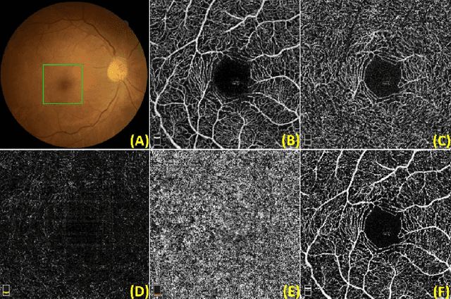
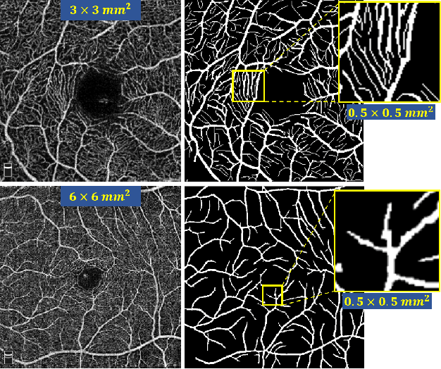
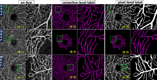
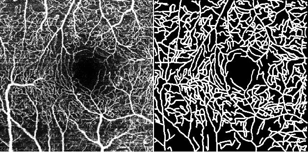
Optical Coherence Tomography Angiography (OCT-A) is a non-invasive imaging technique, and has been increasingly used to image the retinal vasculature at capillary level resolution. However, automated segmentation of retinal vessels in OCT-A has been under-studied due to various challenges such as low capillary visibility and high vessel complexity, despite its significance in understanding many eye-related diseases. In addition, there is no publicly available OCT-A dataset with manually graded vessels for training and validation. To address these issues, for the first time in the field of retinal image analysis we construct a dedicated Retinal OCT-A SEgmentation dataset (ROSE), which consists of 229 OCT-A images with vessel annotations at either centerline-level or pixel level. This dataset has been released for public access to assist researchers in the community in undertaking research in related topics. Secondly, we propose a novel Split-based Coarse-to-Fine vessel segmentation network (SCF-Net), with the ability to detect thick and thin vessels separately. In the SCF-Net, a split-based coarse segmentation (SCS) module is first introduced to produce a preliminary confidence map of vessels, and a split-based refinement (SRN) module is then used to optimize the shape/contour of the retinal microvasculature. Thirdly, we perform a thorough evaluation of the state-of-the-art vessel segmentation models and our SCF-Net on the proposed ROSE dataset. The experimental results demonstrate that our SCF-Net yields better vessel segmentation performance in OCT-A than both traditional methods and other deep learning methods.