 Add to Chrome
 Add to Firefox
 Add to Edge
Time Blindness: Why Video-Language Models Can't See What Humans Can?
May 30, 2025Recent advances in vision-language models (VLMs) have made impressive strides in understanding spatio-temporal relationships in videos. However, when spatial information is obscured, these models struggle to capture purely temporal patterns. We introduce $\textbf{SpookyBench}$, a benchmark where information is encoded solely in temporal sequences of noise-like frames, mirroring natural phenomena from biological signaling to covert communication. Interestingly, while humans can recognize shapes, text, and patterns in these sequences with over 98% accuracy, state-of-the-art VLMs achieve 0% accuracy. This performance gap highlights a critical limitation: an over-reliance on frame-level spatial features and an inability to extract meaning from temporal cues. Furthermore, when trained in data sets with low spatial signal-to-noise ratios (SNR), temporal understanding of models degrades more rapidly than human perception, especially in tasks requiring fine-grained temporal reasoning. Overcoming this limitation will require novel architectures or training paradigms that decouple spatial dependencies from temporal processing. Our systematic analysis shows that this issue persists across model scales and architectures. We release SpookyBench to catalyze research in temporal pattern recognition and bridge the gap between human and machine video understanding. Dataset and code has been made available on our project website: https://timeblindness.github.io/.
3DCoMPaT$^{++}$: An improved Large-scale 3D Vision Dataset for Compositional Recognition
Oct 27, 2023


In this work, we present 3DCoMPaT$^{++}$, a multimodal 2D/3D dataset with 160 million rendered views of more than 10 million stylized 3D shapes carefully annotated at the part-instance level, alongside matching RGB point clouds, 3D textured meshes, depth maps, and segmentation masks. 3DCoMPaT$^{++}$ covers 41 shape categories, 275 fine-grained part categories, and 293 fine-grained material classes that can be compositionally applied to parts of 3D objects. We render a subset of one million stylized shapes from four equally spaced views as well as four randomized views, leading to a total of 160 million renderings. Parts are segmented at the instance level, with coarse-grained and fine-grained semantic levels. We introduce a new task, called Grounded CoMPaT Recognition (GCR), to collectively recognize and ground compositions of materials on parts of 3D objects. Additionally, we report the outcomes of a data challenge organized at CVPR2023, showcasing the winning method's utilization of a modified PointNet$^{++}$ model trained on 6D inputs, and exploring alternative techniques for GCR enhancement. We hope our work will help ease future research on compositional 3D Vision.
Identification of Hemorrhage and Infarct Lesions on Brain CT Images using Deep Learning
Jul 10, 2023Head Non-contrast computed tomography (NCCT) scan remain the preferred primary imaging modality due to their widespread availability and speed. However, the current standard for manual annotations of abnormal brain tissue on head NCCT scans involves significant disadvantages like lack of cutoff standardization and degeneration identification. The recent advancement of deep learning-based computer-aided diagnostic (CAD) models in the multidisciplinary domain has created vast opportunities in neurological medical imaging. Significant literature has been published earlier in the automated identification of brain tissue on different imaging modalities. However, determining Intracranial hemorrhage (ICH) and infarct can be challenging due to image texture, volume size, and scan quality variability. This retrospective validation study evaluated a DL-based algorithm identifying ICH and infarct from head-NCCT scans. The head-NCCT scans dataset was collected consecutively from multiple diagnostic imaging centers across India. The study exhibits the potential and limitations of such DL-based software for introduction in routine workflow in extensive healthcare facilities.
Deep-ASPECTS: A Segmentation-Assisted Model for Stroke Severity Measurement
Mar 17, 2022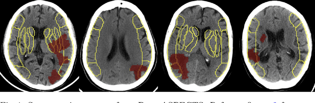
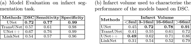
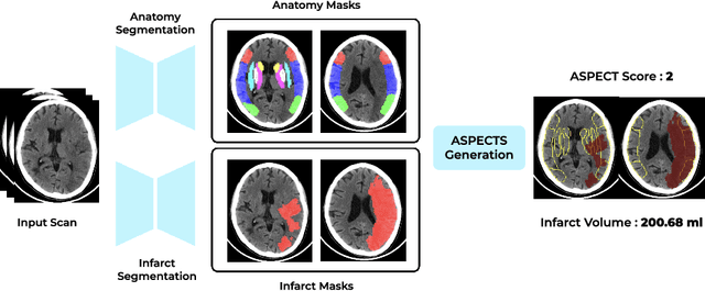
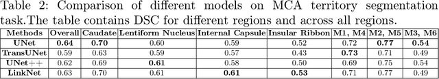
A stroke occurs when an artery in the brain ruptures and bleeds or when the blood supply to the brain is cut off. Blood and oxygen cannot reach the brain's tissues due to the rupture or obstruction resulting in tissue death. The Middle cerebral artery (MCA) is the largest cerebral artery and the most commonly damaged vessel in stroke. The quick onset of a focused neurological deficit caused by interruption of blood flow in the territory supplied by the MCA is known as an MCA stroke. Alberta stroke programme early CT score (ASPECTS) is used to estimate the extent of early ischemic changes in patients with MCA stroke. This study proposes a deep learning-based method to score the CT scan for ASPECTS. Our work has three highlights. First, we propose a novel method for medical image segmentation for stroke detection. Second, we show the effectiveness of AI solution for fully-automated ASPECT scoring with reduced diagnosis time for a given non-contrast CT (NCCT) Scan. Our algorithms show a dice similarity coefficient of 0.64 for the MCA anatomy segmentation and 0.72 for the infarcts segmentation. Lastly, we show that our model's performance is inline with inter-reader variability between radiologists.
Generating Out of Distribution Adversarial Attack using Latent Space Poisoning
Dec 09, 2020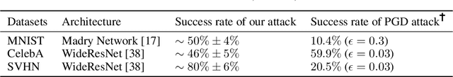
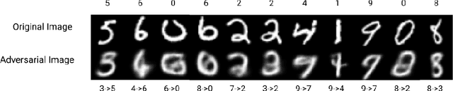
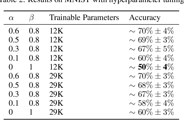
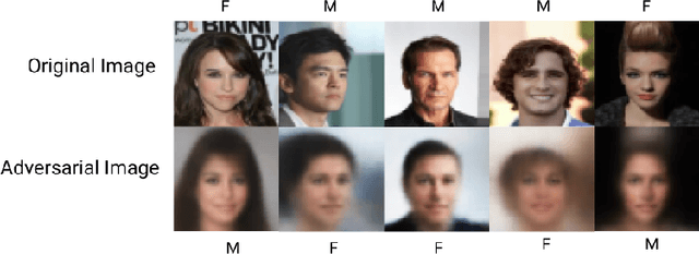
Traditional adversarial attacks rely upon the perturbations generated by gradients from the network which are generally safeguarded by gradient guided search to provide an adversarial counterpart to the network. In this paper, we propose a novel mechanism of generating adversarial examples where the actual image is not corrupted rather its latent space representation is utilized to tamper with the inherent structure of the image while maintaining the perceptual quality intact and to act as legitimate data samples. As opposed to gradient-based attacks, the latent space poisoning exploits the inclination of classifiers to model the independent and identical distribution of the training dataset and tricks it by producing out of distribution samples. We train a disentangled variational autoencoder (beta-VAE) to model the data in latent space and then we add noise perturbations using a class-conditioned distribution function to the latent space under the constraint that it is misclassified to the target label. Our empirical results on MNIST, SVHN, and CelebA dataset validate that the generated adversarial examples can easily fool robust l_0, l_2, l_inf norm classifiers designed using provably robust defense mechanisms.