 Add to Chrome
 Add to Firefox
 Add to Edge
Comparative Evaluation of Digital and Analog Chest Radiographs to Identify Tuberculosis using Deep Learning Model
Jul 27, 2023


Purpose:Chest X-ray (CXR) is an essential tool and one of the most prescribed imaging to detect pulmonary abnormalities, with a yearly estimate of over 2 billion imaging performed worldwide. However, the accurate and timely diagnosis of TB remains an unmet goal. The prevalence of TB is highest in low-middle-income countries, and the requirement of a portable, automated, and reliable solution is required. In this study, we compared the performance of DL-based devices on digital and analog CXR. The evaluated DL-based device can be used in resource-constraint settings. Methods: A total of 10,000 CXR DICOMs(.dcm) and printed photos of the films acquired with three different cellular phones - Samsung S8, iPhone 8, and iPhone XS along with their radiological report were retrospectively collected from various sites across India from April 2020 to March 2021. Results: 10,000 chest X-rays were utilized to evaluate the DL-based device in identifying radiological signs of TB. The AUC of qXR for detecting signs of tuberculosis on the original DICOMs dataset was 0.928 with a sensitivity of 0.841 at a specificity of 0.806. At an optimal threshold, the difference in the AUC of three cellular smartphones with the original DICOMs is 0.024 (2.55%), 0.048 (5.10%), and 0.038 (1.91%). The minimum difference demonstrates the robustness of the DL-based device in identifying radiological signs of TB in both digital and analog CXR.
Identification of Hemorrhage and Infarct Lesions on Brain CT Images using Deep Learning
Jul 10, 2023Head Non-contrast computed tomography (NCCT) scan remain the preferred primary imaging modality due to their widespread availability and speed. However, the current standard for manual annotations of abnormal brain tissue on head NCCT scans involves significant disadvantages like lack of cutoff standardization and degeneration identification. The recent advancement of deep learning-based computer-aided diagnostic (CAD) models in the multidisciplinary domain has created vast opportunities in neurological medical imaging. Significant literature has been published earlier in the automated identification of brain tissue on different imaging modalities. However, determining Intracranial hemorrhage (ICH) and infarct can be challenging due to image texture, volume size, and scan quality variability. This retrospective validation study evaluated a DL-based algorithm identifying ICH and infarct from head-NCCT scans. The head-NCCT scans dataset was collected consecutively from multiple diagnostic imaging centers across India. The study exhibits the potential and limitations of such DL-based software for introduction in routine workflow in extensive healthcare facilities.
StationPlot: A New Non-stationarity Quantification Tool for Detection of Epileptic Seizures
Nov 10, 2018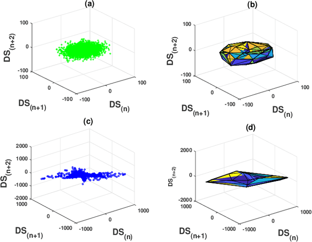
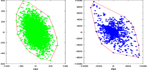
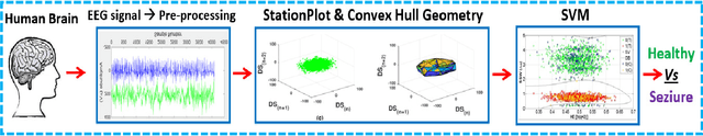
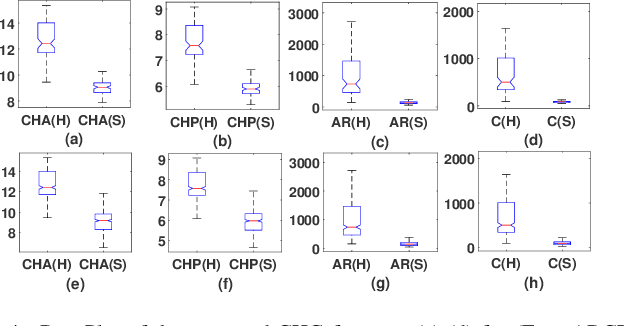
A novel non-stationarity visualization tool known as StationPlot is developed for deciphering the chaotic behavior of a dynamical time series. A family of analytic measures enumerating geometrical aspects of the non-stationarity & degree of variability is formulated by convex hull geometry (CHG) on StationPlot. In the Euclidean space, both trend-stationary (TS) & difference-stationary (DS) perturbations are comprehended by the asymmetric structure of StationPlot's region of interest (ROI). The proposed method is experimentally validated using EEG signals, where it comprehend the relative temporal evolution of neural dynamics & its non-stationary morphology, thereby exemplifying its diagnostic competence for seizure activity (SA) detection. Experimental results & analysis-of-Variance (ANOVA) on the extracted CHG features demonstrates better classification performances as compared to the existing shallow feature based state-of-the-art & validates its efficacy as geometry-rich discriminative descriptors for signal processing applications.
Manifold Learning & Stacked Sparse Autoencoder for Robust Breast Cancer Classification from Histopathological Images
Jun 18, 2018
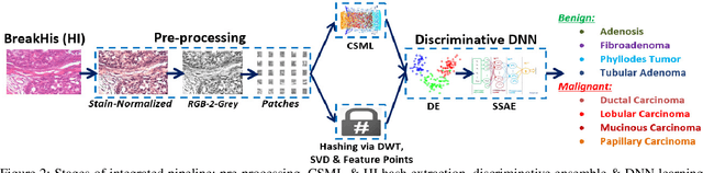

Computer aided diagnosis (CAD) of histopathological images (HI) requires efficient structural representation of the underlying surface tissue convolutions as manifested by the diverse breast cancerous (BC) tissue morphology. In this contribution, HI are modelled as spatially-progressive lower dimensional dynamical patterns embedded in the higher dimensional HI space. Manifold learning on these HI point-cloud is envisaged by LandMark ISOMAP (L-ISOMAP) for isometric feature mapping. The dimensionality reduced L-ISOMAP descriptors are cascaded with stacked sparse autoencoder (SSAE) for learning deep textural feature and tumor malignancy detection thereof. Classification accuracy of 99.4% obtained on publicly available BreaKHis dataset outperforms the state-of-the-art methods and validates it's adequacy as an adjunct tool to clinicians in confirming their diagnosis. Further, employing L-Isomap based manifold embedding, the dimensionality of HI are reduced drastically without significant loss in its discriminating competency. These relieves the GPU requirement for SSAE aided deep learning. Experimental results are discussed in detail.
Classification of histopathological breast cancer images using iterative VMD aided Zernike moments & textural signatures
Jan 15, 2018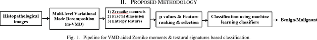
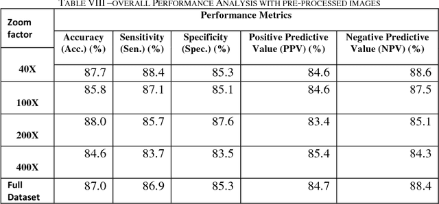
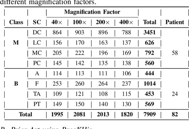
In this paper we present a novel method for an automated diagnosis of breast carcinoma through multilevel iterative variational mode decomposition (VMD) and textural features encompassing Zernaike moments, fractal dimension and entropy features namely, Kapoor entropy, Renyi entropy, Yager entropy features are extracted from VMD components. The proposed method considers the histopathological image as a set of multidimensional spatially-evolving signals. ReliefF algorithm is used to select the discriminatory features and statistically most significant features are fed to squares support vector machine (SVM) for classification. We evaluate the efficiency of the proposed methodology on publicly available Breakhis dataset containing 7,909 breast cancer histological images, collected from 82 patients, of both benign and malignant cases. Experimental results shows the efficacy of the proposed method in outperforming the state of the art while achieving an average classification rates of 89.61% and 88:23% using three-fold and ten-fold cross-validation strategies, respectively. This system can aid the pathologist in accurate and reliable diagnosis of biopsy samples. BreaKHis, a publicly dataset available at http://web.inf.ufpr.br/vri/breast-cancer-database.