 Add to Chrome
 Add to Firefox
 Add to Edge
Empowering Precision Medicine: AI-Driven Schizophrenia Diagnosis via EEG Signals: A Comprehensive Review from 2002-2023
Sep 14, 2023Schizophrenia (SZ) is a prevalent mental disorder characterized by cognitive, emotional, and behavioral changes. Symptoms of SZ include hallucinations, illusions, delusions, lack of motivation, and difficulties in concentration. Diagnosing SZ involves employing various tools, including clinical interviews, physical examinations, psychological evaluations, the Diagnostic and Statistical Manual of Mental Disorders (DSM), and neuroimaging techniques. Electroencephalography (EEG) recording is a significant functional neuroimaging modality that provides valuable insights into brain function during SZ. However, EEG signal analysis poses challenges for neurologists and scientists due to the presence of artifacts, long-term recordings, and the utilization of multiple channels. To address these challenges, researchers have introduced artificial intelligence (AI) techniques, encompassing conventional machine learning (ML) and deep learning (DL) methods, to aid in SZ diagnosis. This study reviews papers focused on SZ diagnosis utilizing EEG signals and AI methods. The introduction section provides a comprehensive explanation of SZ diagnosis methods and intervention techniques. Subsequently, review papers in this field are discussed, followed by an introduction to the AI methods employed for SZ diagnosis and a summary of relevant papers presented in tabular form. Additionally, this study reports on the most significant challenges encountered in SZ diagnosis, as identified through a review of papers in this field. Future directions to overcome these challenges are also addressed. The discussion section examines the specific details of each paper, culminating in the presentation of conclusions and findings.
A Hybrid Deep Spatio-Temporal Attention-Based Model for Parkinson's Disease Diagnosis Using Resting State EEG Signals
Aug 14, 2023


Parkinson's disease (PD), a severe and progressive neurological illness, affects millions of individuals worldwide. For effective treatment and management of PD, an accurate and early diagnosis is crucial. This study presents a deep learning-based model for the diagnosis of PD using resting state electroencephalogram (EEG) signal. The objective of the study is to develop an automated model that can extract complex hidden nonlinear features from EEG and demonstrate its generalizability on unseen data. The model is designed using a hybrid model, consists of convolutional neural network (CNN), bidirectional gated recurrent unit (Bi-GRU), and attention mechanism. The proposed method is evaluated on three public datasets (Uc San Diego Dataset, PRED-CT, and University of Iowa (UI) dataset), with one dataset used for training and the other two for evaluation. The results show that the proposed model can accurately diagnose PD with high performance on both the training and hold-out datasets. The model also performs well even when some part of the input information is missing. The results of this work have significant implications for patient treatment and for ongoing investigations into the early detection of Parkinson's disease. The suggested model holds promise as a non-invasive and reliable technique for PD early detection utilizing resting state EEG.
Full-resolution Lung Nodule Segmentation from Chest X-ray Images using Residual Encoder-Decoder Networks
Jul 13, 2023


Lung cancer is the leading cause of cancer death and early diagnosis is associated with a positive prognosis. Chest X-ray (CXR) provides an inexpensive imaging mode for lung cancer diagnosis. Suspicious nodules are difficult to distinguish from vascular and bone structures using CXR. Computer vision has previously been proposed to assist human radiologists in this task, however, leading studies use down-sampled images and computationally expensive methods with unproven generalization. Instead, this study localizes lung nodules using efficient encoder-decoder neural networks that process full resolution images to avoid any signal loss resulting from down-sampling. Encoder-decoder networks are trained and tested using the JSRT lung nodule dataset. The networks are used to localize lung nodules from an independent external CXR dataset. Sensitivity and false positive rates are measured using an automated framework to eliminate any observer subjectivity. These experiments allow for the determination of the optimal network depth, image resolution and pre-processing pipeline for generalized lung nodule localization. We find that nodule localization is influenced by subtlety, with more subtle nodules being detected in earlier training epochs. Therefore, we propose a novel self-ensemble model from three consecutive epochs centered on the validation optimum. This ensemble achieved a sensitivity of 85% in 10-fold internal testing with false positives of 8 per image. A sensitivity of 81% is achieved at a false positive rate of 6 following morphological false positive reduction. This result is comparable to more computationally complex systems based on linear and spatial filtering, but with a sub-second inference time that is faster than other methods. The proposed algorithm achieved excellent generalization results against an external dataset with sensitivity of 77% at a false positive rate of 7.6.
Remote patient monitoring using artificial intelligence: Current state, applications, and challenges
Jan 19, 2023The adoption of artificial intelligence (AI) in healthcare is growing rapidly. Remote patient monitoring (RPM) is one of the common healthcare applications that assist doctors to monitor patients with chronic or acute illness at remote locations, elderly people in-home care, and even hospitalized patients. The reliability of manual patient monitoring systems depends on staff time management which is dependent on their workload. Conventional patient monitoring involves invasive approaches which require skin contact to monitor health status. This study aims to do a comprehensive review of RPM systems including adopted advanced technologies, AI impact on RPM, challenges and trends in AI-enabled RPM. This review explores the benefits and challenges of patient-centric RPM architectures enabled with Internet of Things wearable devices and sensors using the cloud, fog, edge, and blockchain technologies. The role of AI in RPM ranges from physical activity classification to chronic disease monitoring and vital signs monitoring in emergency settings. This review results show that AI-enabled RPM architectures have transformed healthcare monitoring applications because of their ability to detect early deterioration in patients' health, personalize individual patient health parameter monitoring using federated learning, and learn human behavior patterns using techniques such as reinforcement learning. This review discusses the challenges and trends to adopt AI to RPM systems and implementation issues. The future directions of AI in RPM applications are analyzed based on the challenges and trends
RECOMMED: A Comprehensive Pharmaceutical Recommendation System
Dec 31, 2022


A comprehensive pharmaceutical recommendation system was designed based on the patients and drugs features extracted from Drugs.com and Druglib.com. First, data from these databases were combined, and a dataset of patients and drug information was built. Secondly, the patients and drugs were clustered, and then the recommendation was performed using different ratings provided by patients, and importantly by the knowledge obtained from patients and drug specifications, and considering drug interactions. To the best of our knowledge, we are the first group to consider patients conditions and history in the proposed approach for selecting a specific medicine appropriate for that particular user. Our approach applies artificial intelligence (AI) models for the implementation. Sentiment analysis using natural language processing approaches is employed in pre-processing along with neural network-based methods and recommender system algorithms for modeling the system. In our work, patients conditions and drugs features are used for making two models based on matrix factorization. Then we used drug interaction to filter drugs with severe or mild interactions with other drugs. We developed a deep learning model for recommending drugs by using data from 2304 patients as a training set, and then we used data from 660 patients as our validation set. After that, we used knowledge from critical information about drugs and combined the outcome of the model into a knowledge-based system with the rules obtained from constraints on taking medicine.
Automatic Diagnosis of Myocarditis Disease in Cardiac MRI Modality using Deep Transformers and Explainable Artificial Intelligence
Oct 26, 2022


Myocarditis is among the most important cardiovascular diseases (CVDs), endangering the health of many individuals by damaging the myocardium. Microbes and viruses, such as HIV, play a vital role in myocarditis disease (MCD) incidence. Lack of MCD diagnosis in the early stages is associated with irreversible complications. Cardiac magnetic resonance imaging (CMRI) is highly popular among cardiologists to diagnose CVDs. In this paper, a deep learning (DL) based computer-aided diagnosis system (CADS) is presented for the diagnosis of MCD using CMRI images. The proposed CADS includes dataset, preprocessing, feature extraction, classification, and post-processing steps. First, the Z-Alizadeh dataset was selected for the experiments. The preprocessing step included noise removal, image resizing, and data augmentation (DA). In this step, CutMix, and MixUp techniques were used for the DA. Then, the most recent pre-trained and transformers models were used for feature extraction and classification using CMRI images. Our results show high performance for the detection of MCD using transformer models compared with the pre-trained architectures. Among the DL architectures, Turbulence Neural Transformer (TNT) architecture achieved an accuracy of 99.73% with 10-fold cross-validation strategy. Explainable-based Grad Cam method is used to visualize the MCD suspected areas in CMRI images.
Automated Diagnosis of Cardiovascular Diseases from Cardiac Magnetic Resonance Imaging Using Deep Learning Models: A Review
Oct 26, 2022In recent years, cardiovascular diseases (CVDs) have become one of the leading causes of mortality globally. CVDs appear with minor symptoms and progressively get worse. The majority of people experience symptoms such as exhaustion, shortness of breath, ankle swelling, fluid retention, and other symptoms when starting CVD. Coronary artery disease (CAD), arrhythmia, cardiomyopathy, congenital heart defect (CHD), mitral regurgitation, and angina are the most common CVDs. Clinical methods such as blood tests, electrocardiography (ECG) signals, and medical imaging are the most effective methods used for the detection of CVDs. Among the diagnostic methods, cardiac magnetic resonance imaging (CMR) is increasingly used to diagnose, monitor the disease, plan treatment and predict CVDs. Coupled with all the advantages of CMR data, CVDs diagnosis is challenging for physicians due to many slices of data, low contrast, etc. To address these issues, deep learning (DL) techniques have been employed to the diagnosis of CVDs using CMR data, and much research is currently being conducted in this field. This review provides an overview of the studies performed in CVDs detection using CMR images and DL techniques. The introduction section examined CVDs types, diagnostic methods, and the most important medical imaging techniques. In the following, investigations to detect CVDs using CMR images and the most significant DL methods are presented. Another section discussed the challenges in diagnosing CVDs from CMR data. Next, the discussion section discusses the results of this review, and future work in CVDs diagnosis from CMR images and DL techniques are outlined. The most important findings of this study are presented in the conclusion section.
Automatic Autism Spectrum Disorder Detection Using Artificial Intelligence Methods with MRI Neuroimaging: A Review
Jun 20, 2022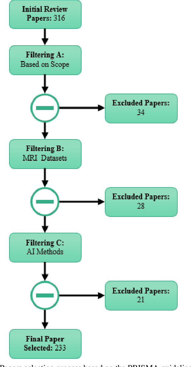
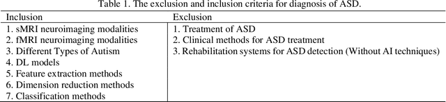
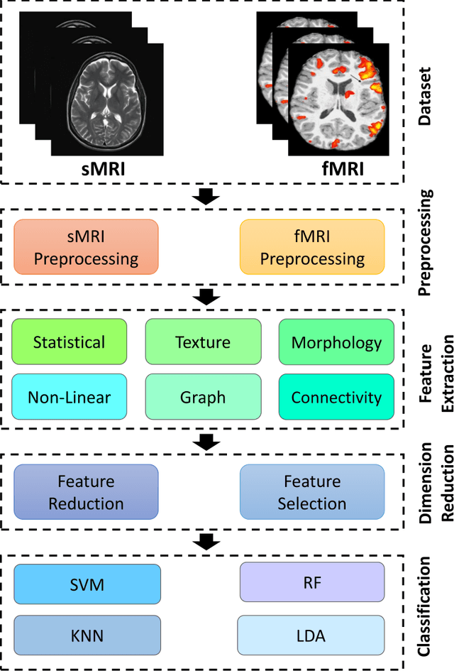
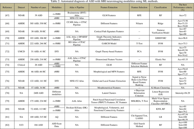
Autism spectrum disorder (ASD) is a brain condition characterized by diverse signs and symptoms that appear in early childhood. ASD is also associated with communication deficits and repetitive behavior in affected individuals. Various ASD detection methods have been developed, including neuroimaging modalities and psychological tests. Among these methods, magnetic resonance imaging (MRI) imaging modalities are of paramount importance to physicians. Clinicians rely on MRI modalities to diagnose ASD accurately. The MRI modalities are non-invasive methods that include functional (fMRI) and structural (sMRI) neuroimaging methods. However, the process of diagnosing ASD with fMRI and sMRI for specialists is often laborious and time-consuming; therefore, several computer-aided design systems (CADS) based on artificial intelligence (AI) have been developed to assist the specialist physicians. Conventional machine learning (ML) and deep learning (DL) are the most popular schemes of AI used for diagnosing ASD. This study aims to review the automated detection of ASD using AI. We review several CADS that have been developed using ML techniques for the automated diagnosis of ASD using MRI modalities. There has been very limited work on the use of DL techniques to develop automated diagnostic models for ASD. A summary of the studies developed using DL is provided in the appendix. Then, the challenges encountered during the automated diagnosis of ASD using MRI and AI techniques are described in detail. Additionally, a graphical comparison of studies using ML and DL to diagnose ASD automatically is discussed. We conclude by suggesting future approaches to detecting ASDs using AI techniques and MRI neuroimaging.
Automatic Diagnosis of Schizophrenia and Attention Deficit Hyperactivity Disorder in rs-fMRI Modality using Convolutional Autoencoder Model and Interval Type-2 Fuzzy Regression
May 31, 2022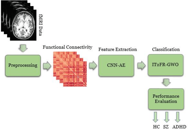
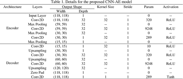
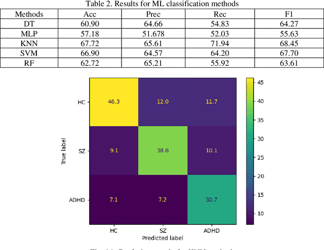
Nowadays, many people worldwide suffer from brain disorders, and their health is in danger. So far, numerous methods have been proposed for the diagnosis of Schizophrenia (SZ) and attention deficit hyperactivity disorder (ADHD), among which functional magnetic resonance imaging (fMRI) modalities are known as a popular method among physicians. This paper presents an SZ and ADHD intelligent detection method of resting-state fMRI (rs-fMRI) modality using a new deep learning (DL) method. The University of California Los Angeles (UCLA) dataset, which contains the rs-fMRI modalities of SZ and ADHD patients, has been used for experiments. The FMRIB software library (FSL) toolbox first performed preprocessing on rs-fMRI data. Then, a convolutional Autoencoder (CNN-AE) model with the proposed number of layers is used to extract features from rs-fMRI data. In the classification step, a new fuzzy method called interval type-2 fuzzy regression (IT2FR) is introduced and then optimized by genetic algorithm (GA), particle swarm optimization (PSO), and gray wolf optimization (GWO) techniques. Also, the results of IT2FR methods are compared with multilayer perceptron (MLP), k-nearest neighbors (KNN), support vector machine (SVM), random forest (RF), decision tree (DT), and adaptive neuro-fuzzy inference system (ANFIS) methods. The experiment results show that the IT2FR method with the GWO optimization algorithm has achieved satisfactory results compared to other classifier methods. Finally, the proposed classification technique was able to provide 72.71% accuracy.
Detection of Epileptic Seizures on EEG Signals Using ANFIS Classifier, Autoencoders and Fuzzy Entropies
Sep 06, 2021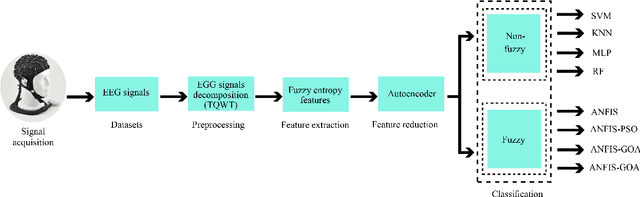
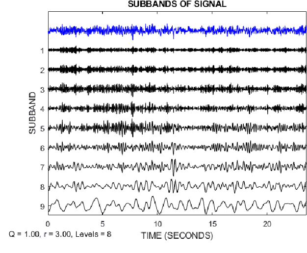
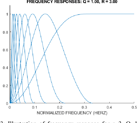

Epilepsy is one of the most crucial neurological disorders, and its early diagnosis will help the clinicians to provide accurate treatment for the patients. The electroencephalogram (EEG) signals are widely used for epileptic seizures detection, which provides specialists with substantial information about the functioning of the brain. In this paper, a novel diagnostic procedure using fuzzy theory and deep learning techniques are introduced. The proposed method is evaluated on the Bonn University dataset with six classification combinations and also on the Freiburg dataset. The tunable-Q wavelet transform (TQWT) is employed to decompose the EEG signals into different sub-bands. In the feature extraction step, 13 different fuzzy entropies are calculated from different sub-bands of TQWT, and their computational complexities are calculated to help researchers choose the best feature sets. In the following, an autoencoder (AE) with six layers is employed for dimensionality reduction. Finally, the standard adaptive neuro-fuzzy inference system (ANFIS), and also its variants with grasshopper optimization algorithm (ANFIS-GOA), particle swarm optimization (ANFIS-PSO), and breeding swarm optimization (ANFIS-BS) methods are used for classification. Using our proposed method, ANFIS-BS method has obtained an accuracy of 99.74% in classifying into two classes and an accuracy of 99.46% in ternary classification on the Bonn dataset and 99.28% on the Freiburg dataset, reaching state-of-the-art performances on both of them.