Juan M. Gorriz
Empowering Precision Medicine: AI-Driven Schizophrenia Diagnosis via EEG Signals: A Comprehensive Review from 2002-2023
Sep 14, 2023Abstract:Schizophrenia (SZ) is a prevalent mental disorder characterized by cognitive, emotional, and behavioral changes. Symptoms of SZ include hallucinations, illusions, delusions, lack of motivation, and difficulties in concentration. Diagnosing SZ involves employing various tools, including clinical interviews, physical examinations, psychological evaluations, the Diagnostic and Statistical Manual of Mental Disorders (DSM), and neuroimaging techniques. Electroencephalography (EEG) recording is a significant functional neuroimaging modality that provides valuable insights into brain function during SZ. However, EEG signal analysis poses challenges for neurologists and scientists due to the presence of artifacts, long-term recordings, and the utilization of multiple channels. To address these challenges, researchers have introduced artificial intelligence (AI) techniques, encompassing conventional machine learning (ML) and deep learning (DL) methods, to aid in SZ diagnosis. This study reviews papers focused on SZ diagnosis utilizing EEG signals and AI methods. The introduction section provides a comprehensive explanation of SZ diagnosis methods and intervention techniques. Subsequently, review papers in this field are discussed, followed by an introduction to the AI methods employed for SZ diagnosis and a summary of relevant papers presented in tabular form. Additionally, this study reports on the most significant challenges encountered in SZ diagnosis, as identified through a review of papers in this field. Future directions to overcome these challenges are also addressed. The discussion section examines the specific details of each paper, culminating in the presentation of conclusions and findings.
Automatic Diagnosis of Myocarditis Disease in Cardiac MRI Modality using Deep Transformers and Explainable Artificial Intelligence
Oct 26, 2022


Abstract:Myocarditis is among the most important cardiovascular diseases (CVDs), endangering the health of many individuals by damaging the myocardium. Microbes and viruses, such as HIV, play a vital role in myocarditis disease (MCD) incidence. Lack of MCD diagnosis in the early stages is associated with irreversible complications. Cardiac magnetic resonance imaging (CMRI) is highly popular among cardiologists to diagnose CVDs. In this paper, a deep learning (DL) based computer-aided diagnosis system (CADS) is presented for the diagnosis of MCD using CMRI images. The proposed CADS includes dataset, preprocessing, feature extraction, classification, and post-processing steps. First, the Z-Alizadeh dataset was selected for the experiments. The preprocessing step included noise removal, image resizing, and data augmentation (DA). In this step, CutMix, and MixUp techniques were used for the DA. Then, the most recent pre-trained and transformers models were used for feature extraction and classification using CMRI images. Our results show high performance for the detection of MCD using transformer models compared with the pre-trained architectures. Among the DL architectures, Turbulence Neural Transformer (TNT) architecture achieved an accuracy of 99.73% with 10-fold cross-validation strategy. Explainable-based Grad Cam method is used to visualize the MCD suspected areas in CMRI images.
Automated Diagnosis of Cardiovascular Diseases from Cardiac Magnetic Resonance Imaging Using Deep Learning Models: A Review
Oct 26, 2022Abstract:In recent years, cardiovascular diseases (CVDs) have become one of the leading causes of mortality globally. CVDs appear with minor symptoms and progressively get worse. The majority of people experience symptoms such as exhaustion, shortness of breath, ankle swelling, fluid retention, and other symptoms when starting CVD. Coronary artery disease (CAD), arrhythmia, cardiomyopathy, congenital heart defect (CHD), mitral regurgitation, and angina are the most common CVDs. Clinical methods such as blood tests, electrocardiography (ECG) signals, and medical imaging are the most effective methods used for the detection of CVDs. Among the diagnostic methods, cardiac magnetic resonance imaging (CMR) is increasingly used to diagnose, monitor the disease, plan treatment and predict CVDs. Coupled with all the advantages of CMR data, CVDs diagnosis is challenging for physicians due to many slices of data, low contrast, etc. To address these issues, deep learning (DL) techniques have been employed to the diagnosis of CVDs using CMR data, and much research is currently being conducted in this field. This review provides an overview of the studies performed in CVDs detection using CMR images and DL techniques. The introduction section examined CVDs types, diagnostic methods, and the most important medical imaging techniques. In the following, investigations to detect CVDs using CMR images and the most significant DL methods are presented. Another section discussed the challenges in diagnosing CVDs from CMR data. Next, the discussion section discusses the results of this review, and future work in CVDs diagnosis from CMR images and DL techniques are outlined. The most important findings of this study are presented in the conclusion section.
Automatic Autism Spectrum Disorder Detection Using Artificial Intelligence Methods with MRI Neuroimaging: A Review
Jun 20, 2022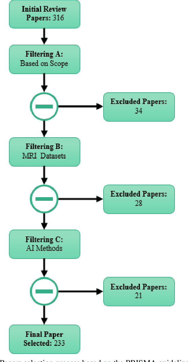

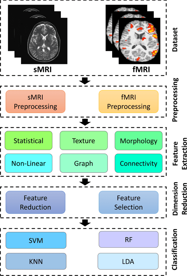
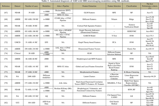
Abstract:Autism spectrum disorder (ASD) is a brain condition characterized by diverse signs and symptoms that appear in early childhood. ASD is also associated with communication deficits and repetitive behavior in affected individuals. Various ASD detection methods have been developed, including neuroimaging modalities and psychological tests. Among these methods, magnetic resonance imaging (MRI) imaging modalities are of paramount importance to physicians. Clinicians rely on MRI modalities to diagnose ASD accurately. The MRI modalities are non-invasive methods that include functional (fMRI) and structural (sMRI) neuroimaging methods. However, the process of diagnosing ASD with fMRI and sMRI for specialists is often laborious and time-consuming; therefore, several computer-aided design systems (CADS) based on artificial intelligence (AI) have been developed to assist the specialist physicians. Conventional machine learning (ML) and deep learning (DL) are the most popular schemes of AI used for diagnosing ASD. This study aims to review the automated detection of ASD using AI. We review several CADS that have been developed using ML techniques for the automated diagnosis of ASD using MRI modalities. There has been very limited work on the use of DL techniques to develop automated diagnostic models for ASD. A summary of the studies developed using DL is provided in the appendix. Then, the challenges encountered during the automated diagnosis of ASD using MRI and AI techniques are described in detail. Additionally, a graphical comparison of studies using ML and DL to diagnose ASD automatically is discussed. We conclude by suggesting future approaches to detecting ASDs using AI techniques and MRI neuroimaging.
Automatic Diagnosis of Schizophrenia and Attention Deficit Hyperactivity Disorder in rs-fMRI Modality using Convolutional Autoencoder Model and Interval Type-2 Fuzzy Regression
May 31, 2022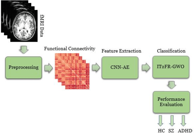
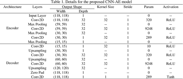
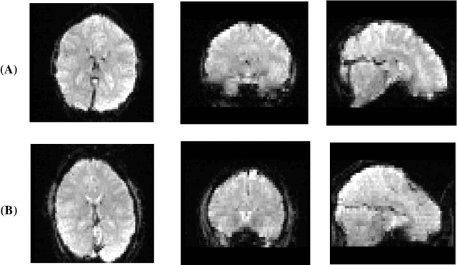
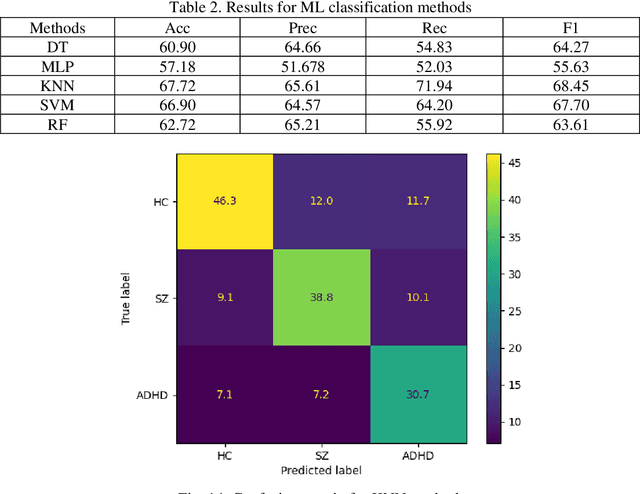
Abstract:Nowadays, many people worldwide suffer from brain disorders, and their health is in danger. So far, numerous methods have been proposed for the diagnosis of Schizophrenia (SZ) and attention deficit hyperactivity disorder (ADHD), among which functional magnetic resonance imaging (fMRI) modalities are known as a popular method among physicians. This paper presents an SZ and ADHD intelligent detection method of resting-state fMRI (rs-fMRI) modality using a new deep learning (DL) method. The University of California Los Angeles (UCLA) dataset, which contains the rs-fMRI modalities of SZ and ADHD patients, has been used for experiments. The FMRIB software library (FSL) toolbox first performed preprocessing on rs-fMRI data. Then, a convolutional Autoencoder (CNN-AE) model with the proposed number of layers is used to extract features from rs-fMRI data. In the classification step, a new fuzzy method called interval type-2 fuzzy regression (IT2FR) is introduced and then optimized by genetic algorithm (GA), particle swarm optimization (PSO), and gray wolf optimization (GWO) techniques. Also, the results of IT2FR methods are compared with multilayer perceptron (MLP), k-nearest neighbors (KNN), support vector machine (SVM), random forest (RF), decision tree (DT), and adaptive neuro-fuzzy inference system (ANFIS) methods. The experiment results show that the IT2FR method with the GWO optimization algorithm has achieved satisfactory results compared to other classifier methods. Finally, the proposed classification technique was able to provide 72.71% accuracy.
What happens in Face during a facial expression? Using data mining techniques to analyze facial expression motion vectors
Sep 12, 2021
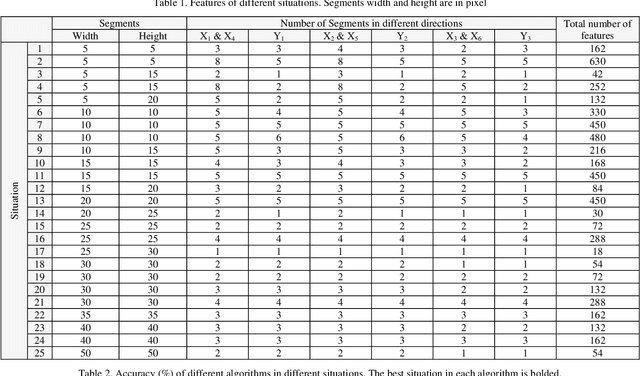

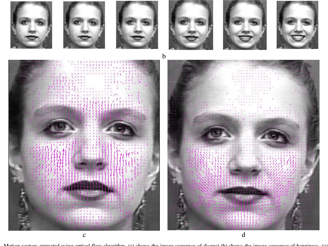
Abstract:One of the most common problems encountered in human-computer interaction is automatic facial expression recognition. Although it is easy for human observer to recognize facial expressions, automatic recognition remains difficult for machines. One of the methods that machines can recognize facial expression is analyzing the changes in face during facial expression presentation. In this paper, optical flow algorithm was used to extract deformation or motion vectors created in the face because of facial expressions. Then, these extracted motion vectors are used to be analyzed. Their positions and directions were exploited for automatic facial expression recognition using different data mining techniques. It means that by employing motion vector features used as our data, facial expressions were recognized. Some of the most state-of-the-art classification algorithms such as C5.0, CRT, QUEST, CHAID, Deep Learning (DL), SVM and Discriminant algorithms were used to classify the extracted motion vectors. Using 10-fold cross validation, their performances were calculated. To compare their performance more precisely, the test was repeated 50 times. Meanwhile, the deformation of face was also analyzed in this research. For example, what exactly happened in each part of face when a person showed fear? Experimental results on Extended Cohen-Kanade (CK+) facial expression dataset demonstrated that the best methods were DL, SVM and C5.0, with the accuracy of 95.3%, 92.8% and 90.2% respectively.
Automatic Diagnosis of Schizophrenia using EEG Signals and CNN-LSTM Models
Sep 02, 2021
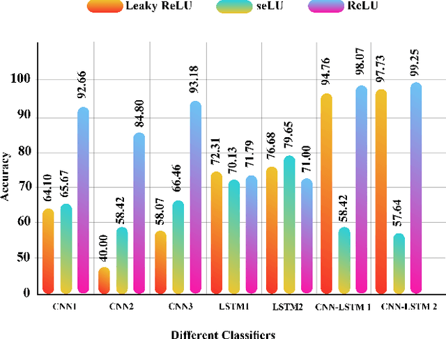
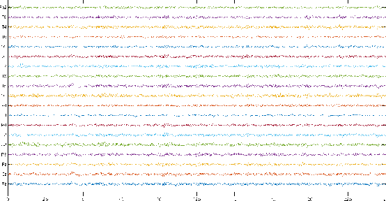
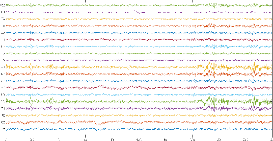
Abstract:Schizophrenia (SZ) is a mental disorder whereby due to the secretion of specific chemicals in the brain, the function of some brain regions is out of balance, leading to the lack of coordination between thoughts, actions, and emotions. This study provides various intelligent Deep Learning (DL)-based methods for automated SZ diagnosis via EEG signals. The obtained results are compared with those of conventional intelligent methods. In order to implement the proposed methods, the dataset of the Institute of Psychiatry and Neurology in Warsaw, Poland, has been used. First, EEG signals are divided into 25-seconds time frames and then were normalized by z-score or norm L2. In the classification step, two different approaches are considered for SZ diagnosis via EEG signals. In this step, the classification of EEG signals is first carried out by conventional DL methods, e.g., KNN, DT, SVM, Bayes, bagging, RF, and ET. Various proposed DL models, including LSTMs, 1D-CNNs, and 1D-CNN-LSTMs, are used in the following. In this step, the DL models were implemented and compared with different activation functions. Among the proposed DL models, the CNN-LSTM architecture has had the best performance. In this architecture, the ReLU activation function and the z-score and L2 combined normalization are used. The proposed CNN-LSTM model has achieved an accuracy percentage of 99.25\%, better than the results of most former studies in this field. It is worth mentioning that in order to perform all simulations, the k-fold cross-validation method with k=5 has been used.
Time Series Forecasting of New Cases and New Deaths Rate for COVID-19 using Deep Learning Methods
Apr 28, 2021
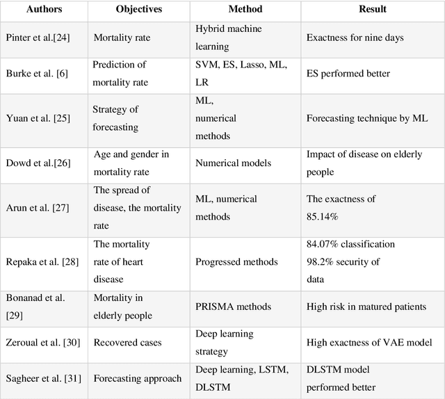
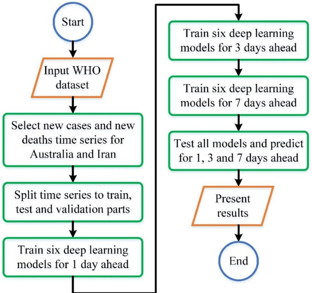

Abstract:Covid-19 has been started in the year 2019 and imposed restrictions in many countries and costs organisations and governments. Predicting the number of new cases and deaths during this period can be a useful step in predicting the costs and facilities required in the future. The purpose of this study is to predict new cases and death rate for seven days ahead. Deep learning methods and statistical analysis model these predictions for 100 days. Six different deep learning methods are examined for the data adopted from the WHO website. Three methods are known as LSTM, Convolutional LSTM, and GRU. The bi-directional mode is then considered for each method to forecast the rate of new cases and new deaths for Australia and Iran countries. This study is novel as it attempts to implement the mentioned three deep learning methods, along with their Bi-directional models, to predict COVID-19 new cases and new death rate time series. All methods are compared, and results are presented. The results are examined in the form of graphs and statistical analyses. The results show that the Bi-directional models have lower error than other models. Several error evaluation metrics are presented to compare all models, and finally, the superiority of Bi-directional methods are determined. The experimental results and statistical test show on datasets to compare the proposed method with other baseline methods. This research could be useful for organisations working against COVID-19 and determining their long-term plans.
CNN AE: Convolution Neural Network combined with Autoencoder approach to detect survival chance of COVID 19 patients
Apr 18, 2021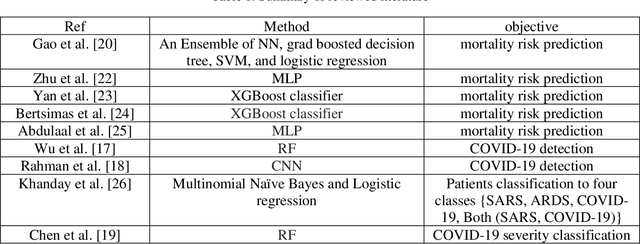
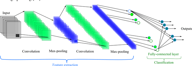
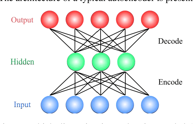
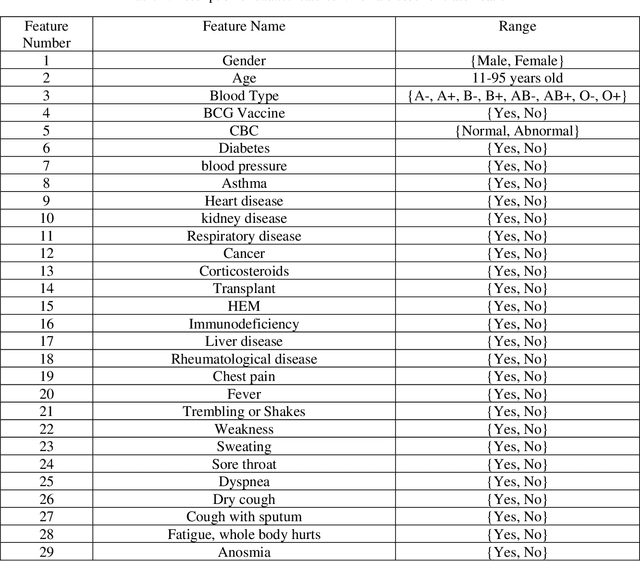
Abstract:In this paper, we propose a novel method named CNN-AE to predict survival chance of COVID-19 patients using a CNN trained on clinical information. To further increase the prediction accuracy, we use the CNN in combination with an autoencoder. Our method is one of the first that aims to predict survival chance of already infected patients. We rely on clinical data to carry out the prediction. The motivation is that the required resources to prepare CT images are expensive and limited compared to the resources required to collect clinical data such as blood pressure, liver disease, etc. We evaluate our method on a publicly available clinical dataset of deceased and recovered patients which we have collected. Careful analysis of the dataset properties is also presented which consists of important features extraction and correlation computation between features. Since most of COVID-19 patients are usually recovered, the number of deceased samples of our dataset is low leading to data imbalance. To remedy this issue, a data augmentation procedure based on autoencoders is proposed. To demonstrate the generality of our augmentation method, we train random forest and Na\"ive Bayes on our dataset with and without augmentation and compare their performance. We also evaluate our method on another dataset for further generality verification. Experimental results reveal the superiority of CNN-AE method compared to the standard CNN as well as other methods such as random forest and Na\"ive Bayes. COVID-19 detection average accuracy of CNN-AE is 96.05% which is higher than CNN average accuracy of 92.49%. To show that clinical data can be used as a reliable dataset for COVID-19 survival chance prediction, CNN-AE is compared with a standard CNN which is trained on CT images.
Uncertainty-Aware Semi-supervised Method using Large Unlabelled and Limited Labeled COVID-19 Data
Feb 12, 2021
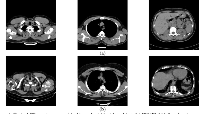
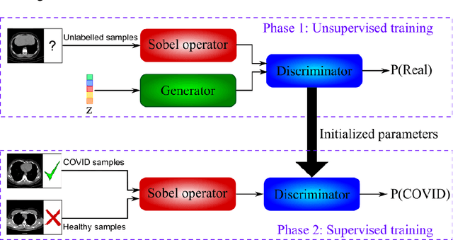
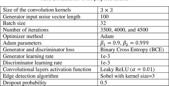
Abstract:The new coronavirus has caused more than 1 million deaths and continues to spread rapidly. This virus targets the lungs, causing respiratory distress which can be mild or severe. The X-ray or computed tomography (CT) images of lungs can reveal whether the patient is infected with COVID-19 or not. Many researchers are trying to improve COVID-19 detection using artificial intelligence. In this paper, relying on Generative Adversarial Networks (GAN), we propose a Semi-supervised Classification using Limited Labelled Data (SCLLD) for automated COVID-19 detection. Our motivation is to develop learning method which can cope with scenarios that preparing labelled data is time consuming or expensive. We further improved the detection accuracy of the proposed method by applying Sobel edge detection. The GAN discriminator output is a probability value which is used for classification in this work. The proposed system is trained using 10,000 CT scans collected from Omid hospital. Also, we validate our system using the public dataset. The proposed method is compared with other state of the art supervised methods such as Gaussian processes. To the best of our knowledge, this is the first time a COVID-19 semi-supervised detection method is presented. Our method is capable of learning from a mixture of limited labelled and unlabelled data where supervised learners fail due to lack of sufficient amount of labelled data. Our semi-supervised training method significantly outperforms the supervised training of Convolutional Neural Network (CNN) in case labelled training data is scarce. Our method has achieved an accuracy of 99.60%, sensitivity of 99.39%, and specificity of 99.80% where CNN (trained supervised) has achieved an accuracy of 69.87%, sensitivity of 94%, and specificity of 46.40%.
 Add to Chrome
 Add to Firefox
 Add to Edge