 Add to Chrome
 Add to Firefox
 Add to Edge
Phenotype discovery of traumatic brain injury segmentations from heterogeneous multi-site data
Nov 05, 2025


Traumatic brain injury (TBI) is intrinsically heterogeneous, and typical clinical outcome measures like the Glasgow Coma Scale complicate this diversity. The large variability in severity and patient outcomes render it difficult to link structural damage to functional deficits. The Federal Interagency Traumatic Brain Injury Research (FITBIR) repository contains large-scale multi-site magnetic resonance imaging data of varying resolutions and acquisition parameters (25 shared studies with 7,693 sessions that have age, sex and TBI status defined - 5,811 TBI and 1,882 controls). To reveal shared pathways of injury of TBI through imaging, we analyzed T1-weighted images from these sessions by first harmonizing to a local dataset and segmenting 132 regions of interest (ROIs) in the brain. After running quality assurance, calculating the volumes of the ROIs, and removing outliers, we calculated the z-scores of volumes for all participants relative to the mean and standard deviation of the controls. We regressed out sex, age, and total brain volume with a multivariate linear regression, and we found significant differences in 37 ROIs between subjects with TBI and controls (p < 0.05 with independent t-tests with false discovery rate correction). We found that differences originated in 1) the brainstem, occipital pole and structures posterior to the orbit, 2) subcortical gray matter and insular cortex, and 3) cerebral and cerebellar white matter using independent component analysis and clustering the component loadings of those with TBI.
Super-Resolution Multi-Contrast Unbiased Eye Atlases With Deep Probabilistic Refinement
Jan 05, 2024


Eye morphology varies significantly across the population, especially for the orbit and optic nerve. These variations limit the feasibility and robustness of generalizing population-wise features of eye organs to an unbiased spatial reference. To tackle these limitations, we propose a process for creating high-resolution unbiased eye atlases. First, to restore spatial details from scans with a low through-plane resolution compared to a high in-plane resolution, we apply a deep learning-based super-resolution algorithm. Then, we generate an initial unbiased reference with an iterative metric-based registration using a small portion of subject scans. We register the remaining scans to this template and refine the template using an unsupervised deep probabilistic approach that generates a more expansive deformation field to enhance the organ boundary alignment. We demonstrate this framework using magnetic resonance images across four different MRI tissue contrasts, generating four atlases in separate spatial alignments. For each tissue contrast, we find a significant improvement in the average Dice score across four labeled regions compared to a standard registration framework consisting of rigid, affine, and deformable transformations. These results highlight the effective alignment of eye organs and boundaries using our proposed process. By combining super-resolution preprocessing and deep probabilistic models, we address the challenge of generating an eye atlas to serve as a standardized reference across a largely variable population.
Joint analysis of structural connectivity and cortical surface features: correlates with mild traumatic brain injury
Dec 15, 2020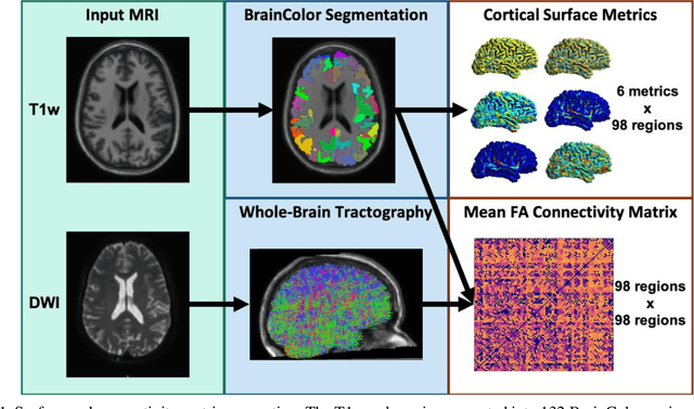
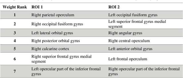
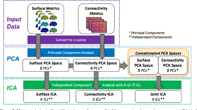
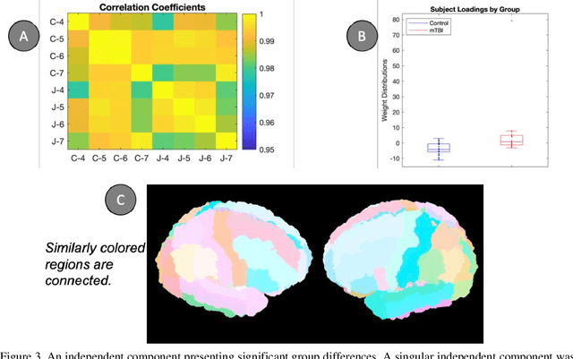
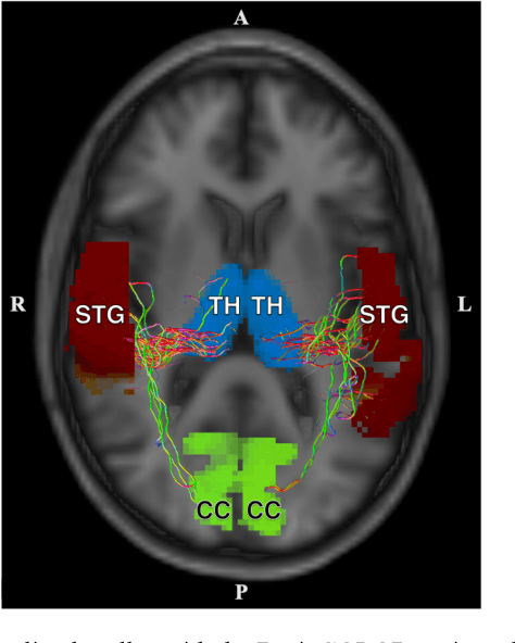
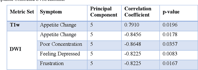
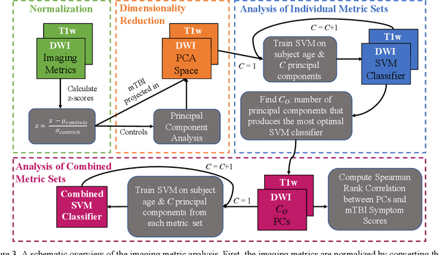
Mild traumatic brain injury (mTBI) is a complex syndrome that affects up to 600 per 100,000 individuals, with a particular concentration among military personnel. About half of all mTBI patients experience a diverse array of chronic symptoms which persist long after the acute injury. Hence, there is an urgent need for better understanding of the white matter and gray matter pathologies associated with mTBI to map which specific brain systems are impacted and identify courses of intervention. Previous works have linked mTBI to disruptions in white matter pathways and cortical surface abnormalities. Herein, we examine these hypothesized links in an exploratory study of joint structural connectivity and cortical surface changes associated with mTBI and its chronic symptoms. Briefly, we consider a cohort of 12 mTBI and 26 control subjects. A set of 588 cortical surface metrics and 4,753 structural connectivity metrics were extracted from cortical surface regions and diffusion weighted magnetic resonance imaging in each subject. Principal component analysis (PCA) was used to reduce the dimensionality of each metric set. We then applied independent component analysis (ICA) both to each PCA space individually and together in a joint ICA approach. We identified a stable independent component across the connectivity-only and joint ICAs which presented significant group differences in subject loadings (p<0.05, corrected). Additionally, we found that two mTBI symptoms, slowed thinking and forgetfulness, were significantly correlated (p<0.05, corrected) with mTBI subject loadings in a surface-only ICA. These surface-only loadings captured an increase in bilateral cortical thickness.
MRI correlates of chronic symptoms in mild traumatic brain injury
Dec 06, 2019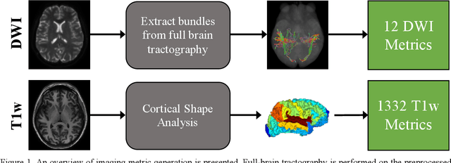
Veterans with mild traumatic brain injury (mTBI) have reported auditory and visual dysfunction that persists beyond the acute incident. The etiology behind these symptoms is difficult to characterize with current clinical imaging. These functional deficits may be caused by shear injury or micro-bleeds, which can be detected with special imaging modalities. We explore these hypotheses in a pilot study of multi-parametric MRI. We extract over 1,000 imaging and clinical metrics and project them to a low-dimensional space, where we can discriminate between healthy controls and patients with mTBI. We also show correlations between the metric representations and patient symptoms.