 Add to Chrome
 Add to Firefox
 Add to Edge
Learning Where and What to Lift for Bi-planar X-ray-to-CT Reconstruction
Aug 18, 2026
X-ray imaging can be approximately modeled as the projection of an underlying volumetric attenuation field, with each measurement recording the accumulated attenuation along a corresponding ray path. Reconstructing a CT volume from only a few X-ray views is therefore severely ill-posed, as the projections collapse depth information and leave 3D locations of anatomical regions and their corresponding intensity distributions highly entangled and ambiguous. We observe that once the spatial organization of anatomical regions is established, estimating their CT intensities becomes substantially more tractable. Motivated by this, we propose LiftXR, an interleaved, geometry-guided framework that explicitly incorporates spatial layout recovery into CT reconstruction. Specifically, a layout lifter first generates a 3D anatomical layout from bi-planar X-rays, providing spatial guidance for an intensity renderer to reconstruct a CT volume. An anatomical parser then performs volumetric perception on the reconstruction, exploiting its spatially resolved boundary and intensity cues to recover a refined anatomical layout. This transition from projection-conditioned layout generation to reconstruction-conditioned anatomical perception allows the parsed layout to provide feedback for region-specific intensity calibration. Extensive experiments on two public datasets demonstrate that LiftXR consistently outperforms recent X-ray-to-CT reconstruction methods, establishing a new state of the art. Moreover, the reconstructed CT achieves superior performance in external downstream segmentation, indicating improved anatomical fidelity. Code will be released.

A Foundation Model for Brain Lesion Segmentation with Mixture of Modality Experts
May 16, 2024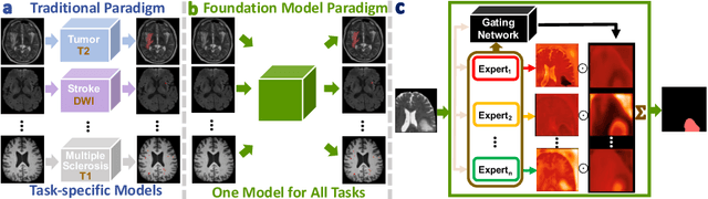
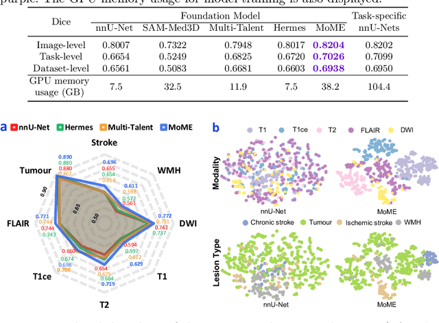
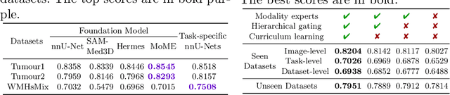
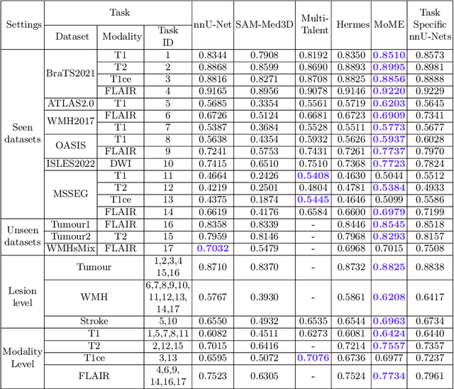
Brain lesion segmentation plays an essential role in neurological research and diagnosis. As brain lesions can be caused by various pathological alterations, different types of brain lesions tend to manifest with different characteristics on different imaging modalities. Due to this complexity, brain lesion segmentation methods are often developed in a task-specific manner. A specific segmentation model is developed for a particular lesion type and imaging modality. However, the use of task-specific models requires predetermination of the lesion type and imaging modality, which complicates their deployment in real-world scenarios. In this work, we propose a universal foundation model for 3D brain lesion segmentation, which can automatically segment different types of brain lesions for input data of various imaging modalities. We formulate a novel Mixture of Modality Experts (MoME) framework with multiple expert networks attending to different imaging modalities. A hierarchical gating network combines the expert predictions and fosters expertise collaboration. Furthermore, we introduce a curriculum learning strategy during training to avoid the degeneration of each expert network and preserve their specialization. We evaluated the proposed method on nine brain lesion datasets, encompassing five imaging modalities and eight lesion types. The results show that our model outperforms state-of-the-art universal models and provides promising generalization to unseen datasets.