 Add to Chrome
 Add to Firefox
 Add to Edge
A WDLoRA-Based Multimodal Generative Framework for Clinically Guided Corneal Confocal Microscopy Image Synthesis in Diabetic Neuropathy
Feb 14, 2026Corneal Confocal Microscopy (CCM) is a sensitive tool for assessing small-fiber damage in Diabetic Peripheral Neuropathy (DPN), yet the development of robust, automated deep learning-based diagnostic models is limited by scarce labelled data and fine-grained variability in corneal nerve morphology. Although Artificial Intelligence (AI)-driven foundation generative models excel at natural image synthesis, they often struggle in medical imaging due to limited domain-specific training, compromising the anatomical fidelity required for clinical analysis. To overcome these limitations, we propose a Weight-Decomposed Low-Rank Adaptation (WDLoRA)-based multimodal generative framework for clinically guided CCM image synthesis. WDLoRA is a parameter-efficient fine-tuning (PEFT) mechanism that decouples magnitude and directional weight updates, enabling foundation generative models to independently learn the orientation (nerve topology) and intensity (stromal contrast) required for medical realism. By jointly conditioning on nerve segmentation masks and disease-specific clinical prompts, the model synthesises anatomically coherent images across the DPN spectrum (Control, T1NoDPN, T1DPN). A comprehensive three-pillar evaluation demonstrates that the proposed framework achieves state-of-the-art visual fidelity (Fréchet Inception Distance (FID): 5.18) and structural integrity (Structural Similarity Index Measure (SSIM): 0.630), significantly outperforming GAN and standard diffusion baselines. Crucially, the synthetic images preserve gold-standard clinical biomarkers and are statistically equivalent to real patient data. When used to train automated diagnostic models, the synthetic dataset improves downstream diagnostic accuracy by 2.1% and segmentation performance by 2.2%, validating the framework's potential to alleviate data bottlenecks in medical AI.
HMSViT: A Hierarchical Masked Self-Supervised Vision Transformer for Corneal Nerve Segmentation and Diabetic Neuropathy Diagnosis
Jun 24, 2025Diabetic Peripheral Neuropathy (DPN) affects nearly half of diabetes patients, requiring early detection. Corneal Confocal Microscopy (CCM) enables non-invasive diagnosis, but automated methods suffer from inefficient feature extraction, reliance on handcrafted priors, and data limitations. We propose HMSViT, a novel Hierarchical Masked Self-Supervised Vision Transformer (HMSViT) designed for corneal nerve segmentation and DPN diagnosis. Unlike existing methods, HMSViT employs pooling-based hierarchical and dual attention mechanisms with absolute positional encoding, enabling efficient multi-scale feature extraction by capturing fine-grained local details in early layers and integrating global context in deeper layers, all at a lower computational cost. A block-masked self supervised learning framework is designed for the HMSViT that reduces reliance on labelled data, enhancing feature robustness, while a multi-scale decoder is used for segmentation and classification by fusing hierarchical features. Experiments on clinical CCM datasets showed HMSViT achieves state-of-the-art performance, with 61.34% mIoU for nerve segmentation and 70.40% diagnostic accuracy, outperforming leading hierarchical models like the Swin Transformer and HiViT by margins of up to 6.39% in segmentation accuracy while using fewer parameters. Detailed ablation studies further reveal that integrating block-masked SSL with hierarchical multi-scale feature extraction substantially enhances performance compared to conventional supervised training. Overall, these comprehensive experiments confirm that HMSViT delivers excellent, robust, and clinically viable results, demonstrating its potential for scalable deployment in real-world diagnostic applications.
A machine learning-based severity prediction tool for diabetic sensorimotor polyneuropathy using Michigan neuropathy screening instrumentations
Mar 28, 2022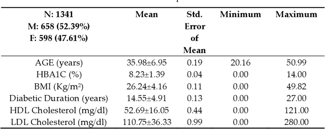
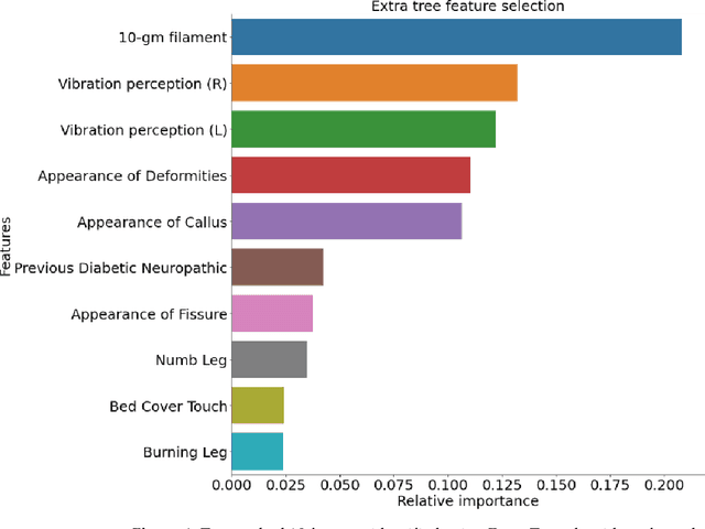
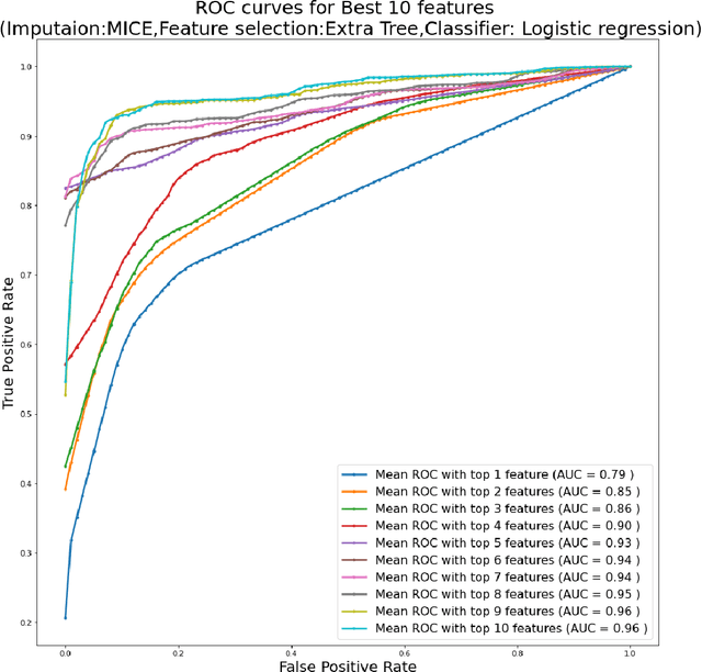
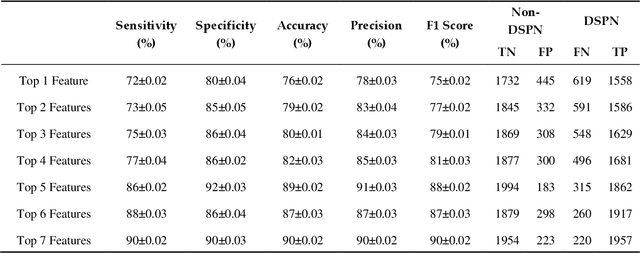
Background: Diabetic Sensorimotor polyneuropathy (DSPN) is a major long-term complication in diabetic patients associated with painful neuropathy, foot ulceration and amputation. The Michigan neuropathy screening instrument (MNSI) is one of the most common screening techniques for DSPN, however, it does not provide any direct severity grading system. Method: For designing and modelling the DSPN severity grading systems for MNSI, 19 years of data from Epidemiology of Diabetes Interventions and Complications (EDIC) clinical trials were used. MNSI variables and patient outcomes were investigated using machine learning tools to identify the features having higher association in DSPN identification. A multivariable logistic regression-based nomogram was generated and validated for DSPN severity grading. Results: The top-7 ranked features from MNSI: 10-gm filament, Vibration perception (R), Vibration perception (L), previous diabetic neuropathy, the appearance of deformities, appearance of callus and appearance of fissure were identified as key features for identifying DSPN using the extra tree model. The area under the curve (AUC) of the nomogram for the internal and external datasets were 0.9421 and 0.946, respectively. From the developed nomogram, the probability of having DSPN was predicted and a DSPN severity scoring system for MNSI was developed from the probability score. The model performance was validated on an independent dataset. Patients were stratified into four severity levels: absent, mild, moderate, and severe using a cut-off value of 10.5, 12.7 and 15 for a DSPN probability less than 50%, 75% to 90%, and above 90%, respectively. Conclusions: This study provides a simple, easy-to-use and reliable algorithm for defining the prognosis and management of patients with DSPN.
A Spatially Constrained Deep Convolutional Neural Network for Nerve Fiber Segmentation in Corneal Confocal Microscopic Images using Inaccurate Annotations
Apr 20, 2020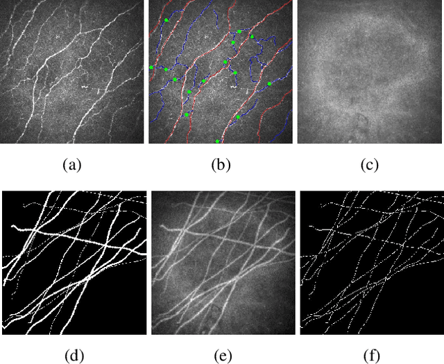
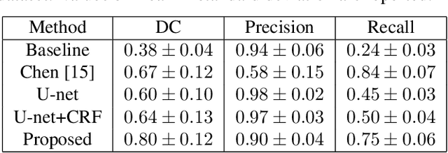
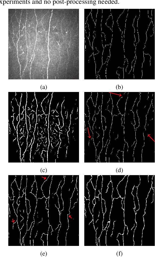
Semantic image segmentation is one of the most important tasks in medical image analysis. Most state-of-the-art deep learning methods require a large number of accurately annotated examples for model training. However, accurate annotation is difficult to obtain especially in medical applications. In this paper, we propose a spatially constrained deep convolutional neural network (DCNN) to achieve smooth and robust image segmentation using inaccurately annotated labels for training. In our proposed method, image segmentation is formulated as a graph optimization problem that is solved by a DCNN model learning process. The cost function to be optimized consists of a unary term that is calculated by cross entropy measurement and a pairwise term that is based on enforcing a local label consistency. The proposed method has been evaluated based on corneal confocal microscopic (CCM) images for nerve fiber segmentation, where accurate annotations are extremely difficult to be obtained. Based on both the quantitative result of a synthetic dataset and qualitative assessment of a real dataset, the proposed method has achieved superior performance in producing high quality segmentation results even with inaccurate labels for training.
* 4 pages, accepted for publication at IEEE International Symposium on Biomedical Imaging (ISBI) 2020