 Add to Chrome
 Add to Firefox
 Add to Edge
Compressive single-pixel imaging via a wavelength-multiplexed spatially incoherent diffractive optical processor
Mar 23, 2026Despite offering high sensitivity, a high signal-to-noise ratio, and a broad spectral range, single-pixel imaging (SPI) is limited by low measurement efficiency and long data-acquisition times. To address this, we propose a wavelength-multiplexed, spatially incoherent diffractive optical processor combined with a compact/shallow digital artificial neural network (ANN) to implement compressive SPI. Specifically, we model the bucket detection process in conventional SPI as a linear intensity transformation with spatially and spectrally varying point-spread functions. This transformation matrix is treated as a learnable parameter and jointly optimized with a shallow digital ANN composed of 2 hidden nonlinear layers. The wavelength-multiplexed diffractive processor is then configured via data-free optimization to approximate this pre-trained transformation matrix; after this optimization, the diffractive processor remains static/fixed. Upon multi-wavelength illumination and diffractive modulation, the target spatial information of the input object is spectrally encoded. A single-pixel detector captures the output spectral power at each illumination band, which is then rapidly decoded by the jointly trained digital ANN to reconstruct the input image. In addition to our numerical analyses demonstrating the feasibility of this approach, we experimentally validated its proof-of-concept using an array of light-emitting diodes (LEDs). Overall, this work demonstrates a computational imaging framework for compressive SPI that can be useful in applications such as biomedical imaging, autonomous devices, and remote sensing.
Pixel Super-Resolved Fluorescence Lifetime Imaging Using Deep Learning
Dec 18, 2025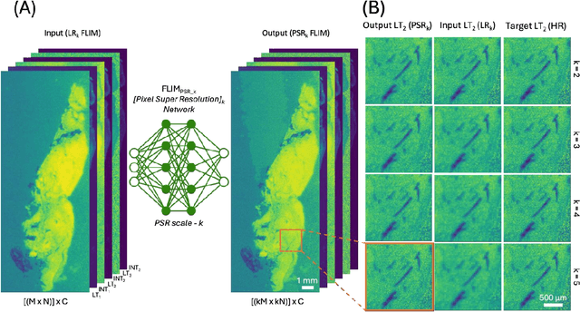
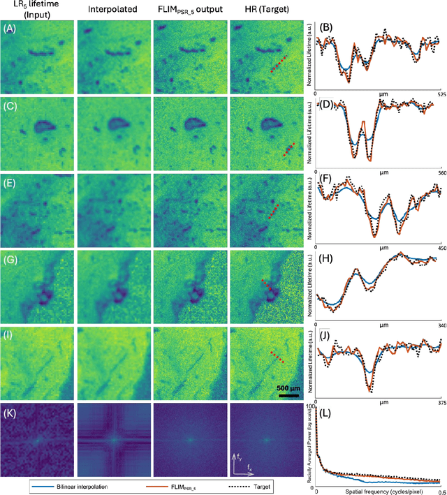
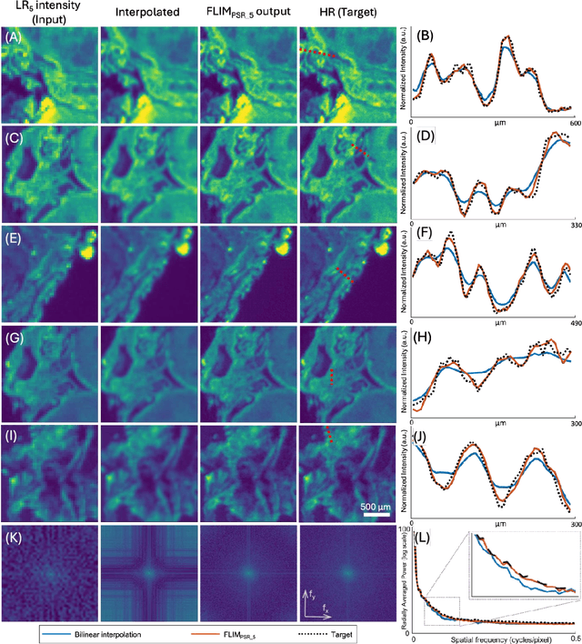
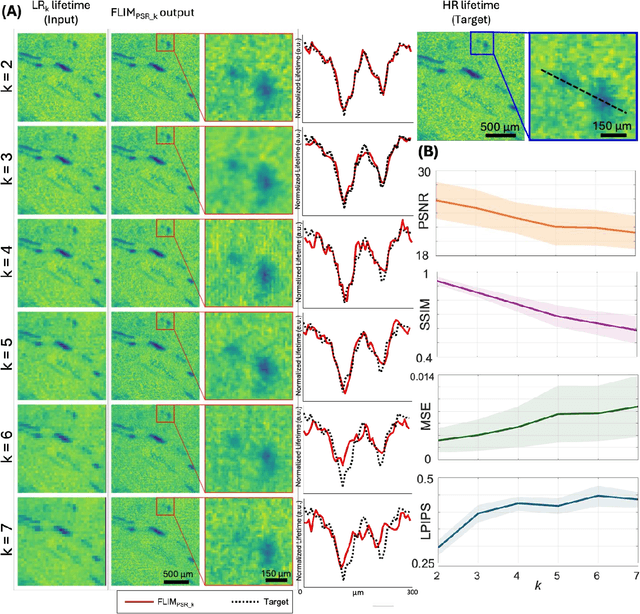
Fluorescence lifetime imaging microscopy (FLIM) is a powerful quantitative technique that provides metabolic and molecular contrast, offering strong translational potential for label-free, real-time diagnostics. However, its clinical adoption remains limited by long pixel dwell times and low signal-to-noise ratio (SNR), which impose a stricter resolution-speed trade-off than conventional optical imaging approaches. Here, we introduce FLIM_PSR_k, a deep learning-based multi-channel pixel super-resolution (PSR) framework that reconstructs high-resolution FLIM images from data acquired with up to a 5-fold increased pixel size. The model is trained using the conditional generative adversarial network (cGAN) framework, which, compared to diffusion model-based alternatives, delivers a more robust PSR reconstruction with substantially shorter inference times, a crucial advantage for practical deployment. FLIM_PSR_k not only enables faster image acquisition but can also alleviate SNR limitations in autofluorescence-based FLIM. Blind testing on held-out patient-derived tumor tissue samples demonstrates that FLIM_PSR_k reliably achieves a super-resolution factor of k = 5, resulting in a 25-fold increase in the space-bandwidth product of the output images and revealing fine architectural features lost in lower-resolution inputs, with statistically significant improvements across various image quality metrics. By increasing FLIM's effective spatial resolution, FLIM_PSR_k advances lifetime imaging toward faster, higher-resolution, and hardware-flexible implementations compatible with low-numerical-aperture and miniaturized platforms, better positioning FLIM for translational applications.
Snapshot multi-spectral imaging through defocusing and a Fourier imager network
Jan 24, 2025


Multi-spectral imaging, which simultaneously captures the spatial and spectral information of a scene, is widely used across diverse fields, including remote sensing, biomedical imaging, and agricultural monitoring. Here, we introduce a snapshot multi-spectral imaging approach employing a standard monochrome image sensor with no additional spectral filters or customized components. Our system leverages the inherent chromatic aberration of wavelength-dependent defocusing as a natural source of physical encoding of multi-spectral information; this encoded image information is rapidly decoded via a deep learning-based multi-spectral Fourier Imager Network (mFIN). We experimentally tested our method with six illumination bands and demonstrated an overall accuracy of 92.98% for predicting the illumination channels at the input and achieved a robust multi-spectral image reconstruction on various test objects. This deep learning-powered framework achieves high-quality multi-spectral image reconstruction using snapshot image acquisition with a monochrome image sensor and could be useful for applications in biomedicine, industrial quality control, and agriculture, among others.
Neural Network-Based Processing and Reconstruction of Compromised Biophotonic Image Data
Mar 21, 2024The integration of deep learning techniques with biophotonic setups has opened new horizons in bioimaging. A compelling trend in this field involves deliberately compromising certain measurement metrics to engineer better bioimaging tools in terms of cost, speed, and form-factor, followed by compensating for the resulting defects through the utilization of deep learning models trained on a large amount of ideal, superior or alternative data. This strategic approach has found increasing popularity due to its potential to enhance various aspects of biophotonic imaging. One of the primary motivations for employing this strategy is the pursuit of higher temporal resolution or increased imaging speed, critical for capturing fine dynamic biological processes. This approach also offers the prospect of simplifying hardware requirements/complexities, thereby making advanced imaging standards more accessible in terms of cost and/or size. This article provides an in-depth review of the diverse measurement aspects that researchers intentionally impair in their biophotonic setups, including the point spread function, signal-to-noise ratio, sampling density, and pixel resolution. By deliberately compromising these metrics, researchers aim to not only recuperate them through the application of deep learning networks, but also bolster in return other crucial parameters, such as the field-of-view, depth-of-field, and space-bandwidth product. Here, we discuss various biophotonic methods that have successfully employed this strategic approach. These techniques span broad applications and showcase the versatility and effectiveness of deep learning in the context of compromised biophotonic data. Finally, by offering our perspectives on the future possibilities of this rapidly evolving concept, we hope to motivate our readers to explore novel ways of balancing hardware compromises with compensation via AI.
Label- and slide-free tissue histology using 3D epi-mode quantitative phase imaging and virtual H&E staining
Jun 01, 2023


Histological staining of tissue biopsies, especially hematoxylin and eosin (H&E) staining, serves as the benchmark for disease diagnosis and comprehensive clinical assessment of tissue. However, the process is laborious and time-consuming, often limiting its usage in crucial applications such as surgical margin assessment. To address these challenges, we combine an emerging 3D quantitative phase imaging technology, termed quantitative oblique back illumination microscopy (qOBM), with an unsupervised generative adversarial network pipeline to map qOBM phase images of unaltered thick tissues (i.e., label- and slide-free) to virtually stained H&E-like (vH&E) images. We demonstrate that the approach achieves high-fidelity conversions to H&E with subcellular detail using fresh tissue specimens from mouse liver, rat gliosarcoma, and human gliomas. We also show that the framework directly enables additional capabilities such as H&E-like contrast for volumetric imaging. The quality and fidelity of the vH&E images are validated using both a neural network classifier trained on real H&E images and tested on virtual H&E images, and a user study with neuropathologists. Given its simple and low-cost embodiment and ability to provide real-time feedback in vivo, this deep learning-enabled qOBM approach could enable new workflows for histopathology with the potential to significantly save time, labor, and costs in cancer screening, detection, treatment guidance, and more.