 Add to Chrome
 Add to Firefox
 Add to Edge
Spinal-Multiple-Myeloma-SEG: A Dual-Energy CT Dataset Extended with Trabecular Bone Segmentation of Lumbar Vertebrae
Aug 02, 2026
We present an extension of the publicly available \textit{Spinal-Multiple-Myeloma-SEG} dataset, a dual-energy CT imaging resource for multiple myeloma research. The purpose of this dataset is to enable voxel-wise analysis of vertebral bone microstructure by adding expert-validated segmentation of the trabecular compartment of lumbar vertebrae. The dataset consists of 72 dual-energy CT examinations from 67 adult patients (mean age 66 years, range 48--85; 36\% female), acquired retrospectively using a dual-layer dual-energy CT system. It includes conventional CT, virtual monoenergetic images, and calcium-suppressed reconstructions, along with structured clinical metadata. The data are provided in DICOM format, while segmentation masks are available in both NIfTI and DICOM-SEG formats. The primary intended applications include trabecular bone segmentation, quantitative bone mineral density-related analysis, and development of deep learning models for vertebral anatomy and disease-affected bone structures in multiple myeloma. The dataset supports both segmentation and multimodal learning tasks in pathological and non-pathological bone. Initial trabecular segmentation masks were generated using a pretrained nnU-Net model and subsequently refined through manual expert correction and radiological quality control, ensuring anatomical consistency. The original dataset is publicly available via TCIA (\href{https://doi.org/10.7937/k4qv-hh78}{https://doi.org/10.7937/k4qv-hh78}), while the trabecular segmentation extension (Version 2) is released through Zenodo (\href{https://doi.org/10.5281/zenodo.21628232}{https://doi.org/10.5281/zenodo.21628232}) under the CC BY 4.0 license. The Zenodo release provides immediate public access to the segmentation masks and will be additionally incorporated into the official TCIA collection after completion of the curation process.

A Unified Framework for Foreground and Anonymization Area Segmentation in CT and MRI Data
Jan 08, 2025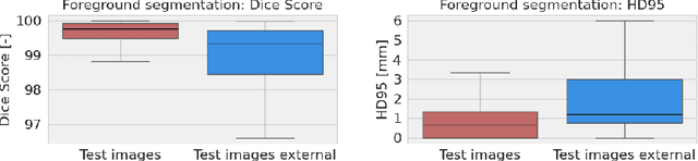

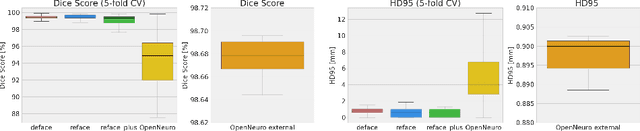
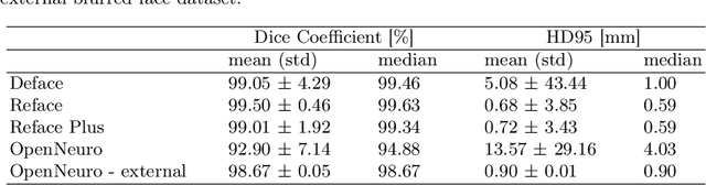
This study presents an open-source toolkit to address critical challenges in preprocessing data for self-supervised learning (SSL) for 3D medical imaging, focusing on data privacy and computational efficiency. The toolkit comprises two main components: a segmentation network that delineates foreground regions to optimize data sampling and thus reduce training time, and a segmentation network that identifies anonymized regions, preventing erroneous supervision in reconstruction-based SSL methods. Experimental results demonstrate high robustness, with mean Dice scores exceeding 98.5 across all anonymization methods and surpassing 99.5 for foreground segmentation tasks, highlighting the efficacy of the toolkit in supporting SSL applications in 3D medical imaging for both CT and MRI images. The weights and code is available at https://github.com/MIC-DKFZ/Foreground-and-Anonymization-Area-Segmentation.
An OpenMind for 3D medical vision self-supervised learning
Dec 22, 2024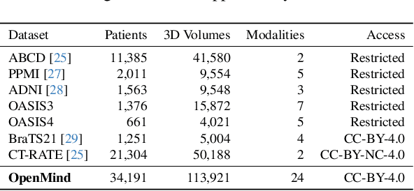
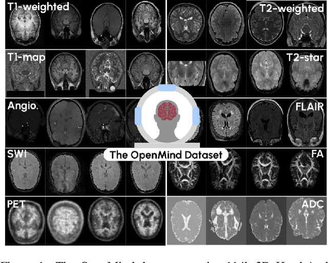
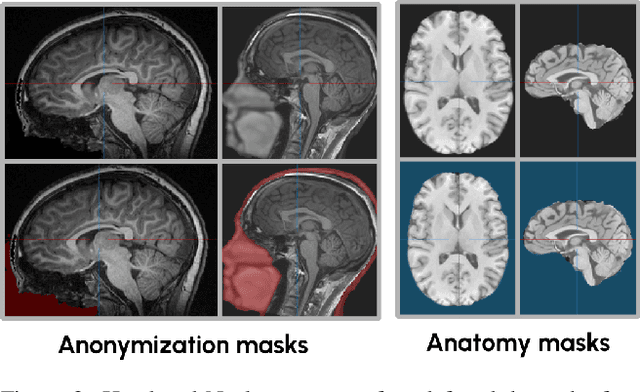
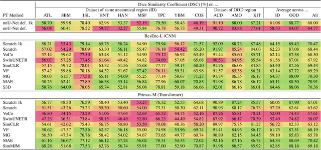
The field of 3D medical vision self-supervised learning lacks consistency and standardization. While many methods have been developed it is impossible to identify the current state-of-the-art, due to i) varying and small pre-training datasets, ii) varying architectures, and iii) being evaluated on differing downstream datasets. In this paper we bring clarity to this field and lay the foundation for further method advancements: We a) publish the largest publicly available pre-training dataset comprising 114k 3D brain MRI volumes and b) benchmark existing SSL methods under common architectures and c) provide the code of our framework publicly to facilitate rapid adoption and reproduction. This pre-print \textit{only describes} the dataset contribution (a); Data, benchmark, and codebase will be made available shortly.