 Add to Chrome
 Add to Firefox
 Add to Edge
Spinal-Multiple-Myeloma-SEG: A Dual-Energy CT Dataset Extended with Trabecular Bone Segmentation of Lumbar Vertebrae
Aug 02, 2026
We present an extension of the publicly available \textit{Spinal-Multiple-Myeloma-SEG} dataset, a dual-energy CT imaging resource for multiple myeloma research. The purpose of this dataset is to enable voxel-wise analysis of vertebral bone microstructure by adding expert-validated segmentation of the trabecular compartment of lumbar vertebrae. The dataset consists of 72 dual-energy CT examinations from 67 adult patients (mean age 66 years, range 48--85; 36\% female), acquired retrospectively using a dual-layer dual-energy CT system. It includes conventional CT, virtual monoenergetic images, and calcium-suppressed reconstructions, along with structured clinical metadata. The data are provided in DICOM format, while segmentation masks are available in both NIfTI and DICOM-SEG formats. The primary intended applications include trabecular bone segmentation, quantitative bone mineral density-related analysis, and development of deep learning models for vertebral anatomy and disease-affected bone structures in multiple myeloma. The dataset supports both segmentation and multimodal learning tasks in pathological and non-pathological bone. Initial trabecular segmentation masks were generated using a pretrained nnU-Net model and subsequently refined through manual expert correction and radiological quality control, ensuring anatomical consistency. The original dataset is publicly available via TCIA (\href{https://doi.org/10.7937/k4qv-hh78}{https://doi.org/10.7937/k4qv-hh78}), while the trabecular segmentation extension (Version 2) is released through Zenodo (\href{https://doi.org/10.5281/zenodo.21628232}{https://doi.org/10.5281/zenodo.21628232}) under the CC BY 4.0 license. The Zenodo release provides immediate public access to the segmentation masks and will be additionally incorporated into the official TCIA collection after completion of the curation process.

Weakly supervised deep learning-based intracranial hemorrhage localization
May 03, 2021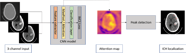
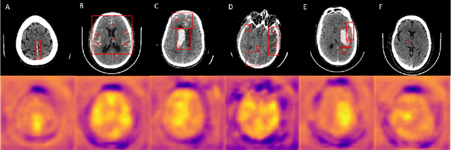
Intracranial hemorrhage is a life-threatening disease, which requires fast medical intervention. Owing to the duration of data annotation, head CT images are usually available only with slice-level labeling. This paper presents a weakly supervised method of precise hemorrhage localization in axial slices using only position-free labels, which is based on multiple instance learning. An algorithm is introduced that generates hemorrhage likelihood maps and finds the coordinates of bleeding. The Dice coefficient of 58.08 % is achieved on data from a publicly available dataset.
Localization and classification of intracranialhemorrhages in CT data
Sep 07, 2020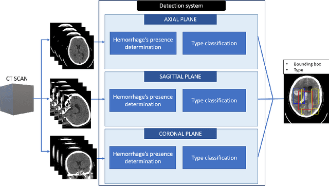
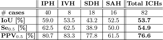
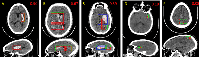
Intracranial hemorrhages (ICHs) are life-threatening brain injures with a relatively high incidence. In this paper, the automatic algorithm for the detection and classification of ICHs, including localization, is present. The set of binary convolutional neural network-based classifiers with a designed cascade-parallel architecture is used. This automatic system may lead to a distinct decrease in the diagnostic process's duration in acute cases. An average Jaccard coefficient of 53.7 % is achieved on the data from the publicly available head CT dataset CQ500.
A Tool for Automatic Estimation of Patient Position in Spinal CT Data
Jun 27, 2020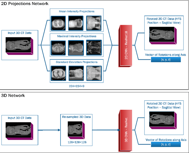
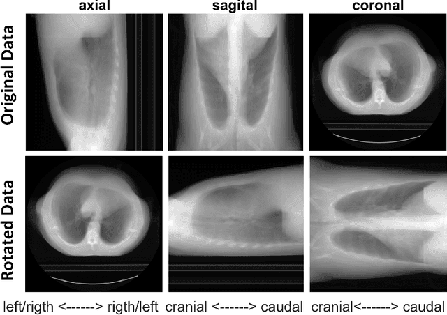
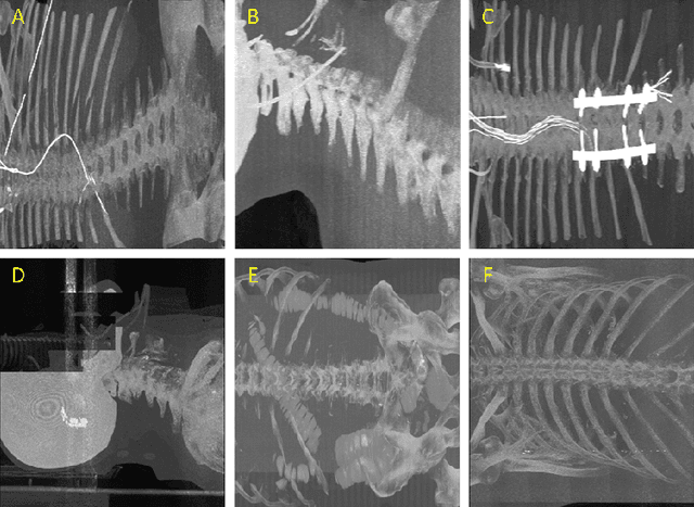
Much of the recently available research and challenge data lack the meta-data containing any information about the patient position. This paper presents a tool for automatic rotation of CT data into a standardized (HFS) patient position. The proposed method is based on the prediction of rotation angle with CNN, and it achieved nearly perfect results with an accuracy of 99.55 %. We provide implementations with easy to use an example for both Matlab and Python (PyTorch), which can be used, for example, for automatic rotation correction of VerSe2020 challenge data.