 Add to Chrome
 Add to Firefox
 Add to Edge
Increasing ultrasound field-of-view with reduced element count arrays containing large elements
Feb 16, 2026Several applications of medical ultrasound can benefit from a larger imaging field of view (FOV). This study is aimed at increasing the FOV of linear array probes by increasing the element size rather than the element count. To investigate larger FOV, this study used coupled elements to imitate a larger element size. The effects of coupling on array beam patterns are examined with Fourier transforms of elements. The effects of coupling on resolution, contrast, and speckle signal-to-noise ratio are examined through phantom images and in-vivo images of a rabbit tumor reconstructed with plane-wave compounding. Furthermore, a positioning system was used to acquire data from a virtual large aperture with 120 mm FOV and 128 elements, collected in sections with a single probe. This study also investigates the Null Subtraction Imaging (NSI), Sign Coherence Factor (SCF), and Minimum Variance (MV) beamformers for regaining resolution lost by an increased F-number with large elements. The MV beamformer, while the most computationally expensive, was best for improving resolution without increasing speckle variance, decreasing Full-Width at Half-Max (FWHM) estimates of wire targets from 0.78 mm with DAS on a 2.5 wavelength element size to 0.54 mm with MV on a 5 wavelength element size.
Large elements and advanced beamformers for increased field of view in 2-D ultrasound matrix arrays
Feb 16, 2026Three-dimensional (3D) ultrasound promises various medical applications for abdominal, obstetrics, and cardiovascular imaging. However, ultrasound matrix arrays have extremely high element counts limiting their field of view (FOV). This work seeks to demonstrate an increased field-of-view using a reduced element count array design. The approach is to increase the element size and use advanced beamformers to maintain image quality. The delay and sum (DAS), Null Subtraction Imaging (NSI), directional coherence factor (DCF), and Minimum Variance (MV) beamformers were compared. K-wave simulations of the 3D point-spread functions (PSF) of NSI, DCF, and MV display reduced side lobes and narrowed main lobes compared to DAS. Experiments were conducted using a multiplexed 1024-element matrix array on a Verasonics 256 system. Elements were electronically coupled to imitate a larger pitch and element size. Then, a virtual large aperture was created by using a positioning system to collect data in sections with the matrix array. High-quality images were obtained using a coupling factor of two, doubling the FOV while maintaining the same element count in the virtual large aperture as the original matrix array. The NSI beamformer demonstrated the best resolution performance in simulations and on the large aperture, maintaining the same resolution as uncoupled DAS for coupling factors up to 4. Our results demonstrate how larger matrix arrays could be constructed with larger elements, with resolution maintained by advanced beamformers.
Label-free Super-Resolution Microvessel Color Flow Imaging with Ultrasound
May 27, 2025

We present phase subtraction imaging (PSI), a new spatial-temporal beamforming method that enables micrometer level resolution imaging of microvessels in live animals without labels, which are microbubbles in ultrasound super-resolution imaging. Subtraction of relative phase differences between consecutive frames beamformed with mismatched apodizations is used in PSI to overcome the diffraction limit. We validated this method by imaging both the mouse brain and rabbit kidney using different ultrasound probes and scanning machines.
The Art of the Steal: Purloining Deep Learning Models Developed for an Ultrasound Scanner to a Competitor Machine
Jul 03, 2024A transfer function approach has recently proven effective for calibrating deep learning (DL) algorithms in quantitative ultrasound (QUS), addressing data shifts at both the acquisition and machine levels. Expanding on this approach, we develop a strategy to 'steal' the functionality of a DL model from one ultrasound machine and implement it on another, in the context of QUS. This demonstrates the ease with which the functionality of a DL model can be transferred between machines, highlighting the security risks associated with deploying such models in a commercial scanner for clinical use. The proposed method is a black-box unsupervised domain adaptation technique that integrates the transfer function approach with an iterative schema. It does not utilize any information related to model internals of the victim machine but it solely relies on the availability of input-output interface. Additionally, we assume the availability of unlabelled data from the testing machine, i.e., the perpetrator machine. This scenario could become commonplace as companies begin deploying their DL functionalities for clinical use. Competing companies might acquire the victim machine and, through the input-output interface, replicate the functionality onto their own machines. In the experiments, we used a SonixOne and a Verasonics machine. The victim model was trained on SonixOne data, and its functionality was then transferred to the Verasonics machine. The proposed method successfully transferred the functionality to the Verasonics machine, achieving a remarkable 98\% classification accuracy in a binary decision task. This study underscores the need to establish security measures prior to deploying DL models in clinical settings.
Machine-to-Machine Transfer Function in Deep Learning-Based Quantitative Ultrasound
Nov 27, 2023A Transfer Function approach was recently demonstrated to mitigate data mismatches at the acquisition level for a single ultrasound scanner in deep learning (DL) based quantitative ultrasound (QUS). As a natural progression, we further investigate the transfer function approach and introduce a Machine-to-Machine (M2M) Transfer Function, which possesses the ability to mitigate data mismatches at a machine level, i.e., mismatches between two scanners over the same frequency band. This ability opens the door to unprecedented opportunities for reducing DL model development costs, enabling the combination of data from multiple sources or scanners, or facilitating the transfer of DL models between machines with ease. We tested the proposed method utilizing a SonixOne machine and a Verasonics machine. In the experiments, we used a L9-4 array and conducted two types of acquisitions to obtain calibration data: stable and free-hand, using two different calibration phantoms. Without the proposed calibration method, the mean classification accuracy when applying a model on data acquired from one system to data acquired from another system was approximately 50%, and the mean AUC was about 0.40. With the proposed method, mean accuracy increased to approximately 90%, and the AUC rose to the 0.99. Additional observations include that shifts in statistics for the z-score normalization had a significant impact on performance. Furthermore, the choice of the calibration phantom played an important role in the proposed method. Additionally, robust implementation inspired by Wiener filtering provided an effective method for transferring the domain from one machine to another machine, and it can succeed using just a single calibration view without the need for multiple independent calibration frames.
A Radiological Clip Design Using Ultrasound Identification to Improve Localization
Aug 30, 2023


Objective: We demonstrate the use of ultrasound to receive an acoustic signal transmitted from a radiological clip designed from a custom circuit. This signal encodes an identification number and is localized and identified wirelessly by the ultrasound imaging system. Methods: We designed and constructed the test platform with a Teensy 4.0 microcontroller core to detect ultrasonic imaging pulses received by a transducer embedded in a phantom, which acted as the radiological clip. Ultrasound identification (USID) signals were generated and transmitted as a result. The phantom and clip were imaged using an ultrasonic array (Philips L7-4) connected to a Verasonics Vantage 128 system operating in pulse inversion (PI) mode. Cross-correlations were performed to localize and identify the code sequences in the PI images. Results: USID signals were detected and visualized on B-mode images of the phantoms with up to sub-millimeter localization accuracy. The average detection rate across 1,600 frames of ultrasound data was 94.6%. Tested ID values exhibited differences in detection rates. Conclusion: The USID clip produced identifiable, distinguishable, and localizable signals when imaged. Significance: Radiological clips are used to mark breast cancer being treated by neoadjuvant chemotherapy (NAC) via implant in or near treated lesions. As NAC progresses, available marking clips can lose visibility in ultrasound, the imaging modality of choice for monitoring NAC-treated lesions. By transmitting an active signal, more accurate and reliable ultrasound localization of these clips could be achieved and multiple clips with different ID values could be imaged in the same field of view.
High-resolution Power Doppler Using Null Subtraction Imaging
Jan 09, 2023


To improve the spatial resolution of power Doppler (PD) imaging, we explored null subtraction imaging (NSI) as an alternative beamforming technique to delay-and-sum (DAS). NSI is a nonlinear beamforming approach that uses three different apodizations on receive and incoherently sums the beamformed envelopes. NSI uses a null in the beam pattern to improve the lateral resolution, which we apply here for improving PD spatial resolution both with and without contrast microbubbles. In this study, we used NSI with singular value decomposition (SVD)-based clutter filtering and noise equalization to generate high-resolution PD images. An element sensitivity correction scheme was also performed to further improve the image quality of PD images using NSI. First, a microbubble trace experiment was performed to quantitatively evaluate the performance of NSI based PD. Then, both contrast-enhanced and contrast free ultrasound data were collected from a rat brain. Higher spatial resolution and image quality were observed from the NSI-based PD microvessel images compared to microvessel images generated by traditional DAS-based beamforming.
Grating lobe reduction in plane wave imaging with angular compounding using subtraction of coherent signals
Oct 24, 2022

Plane wave imaging (PWI) with angular compounding has gained in popularity over recent years because it provides high frame rates and good image properties. However, most linear arrays used in clinical practice have a pitch that is equal to than the wavelength of ultrasound. Hence, the presence of grating lobes is a concern for PWI using multiple transmit angles. The presence of grating lobes produces clutter in images and reduces the ability to observe tissue contrast. Techniques to reduce or eliminate the presence of grating lobes for PWI using multiple angles will result in improved image quality. Null subtraction imaging (NSI) is a nonlinear beamforming technique that has been explored for improving the lateral resolution of ultrasonic imaging. However, the apodization scheme used in NSI also eliminates or greatly reduces the presence of grating lobes. Imaging tasks using NSI were evaluated in simulations and physical experiments involving tissue-mimicking phantoms and rat tumors in vivo. Images created with NSI were compared with images created using traditional delay and sum (DAS) with Hann apodization and images created using a generalized coherence factor (GCF). NSI was observed to greatly reduce the presence of grating lobes in ultrasonic images, compared to DAS with Hann and GCF, while maintaining spatial resolution and contrast in the images. Therefore, NSI can provide a novel means of creating images using PWI with multiple steering angles on clinically available linear arrays while reducing the adverse effects associated with grating lobes.
Through Tissue Ultra-high-definition Video Transmission Using an Ultrasound Communication Channel
Oct 18, 2022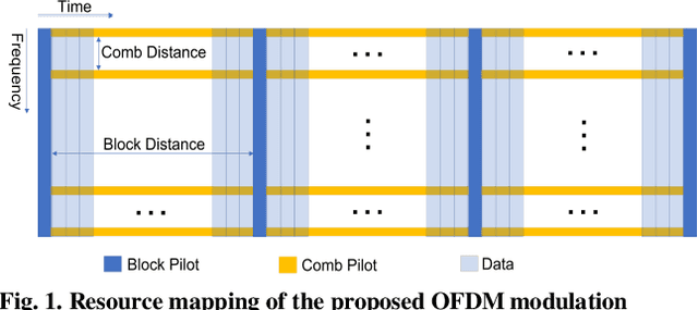
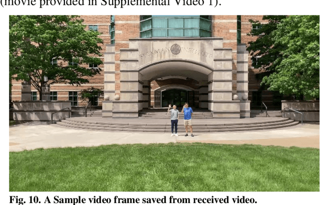
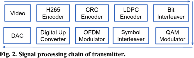
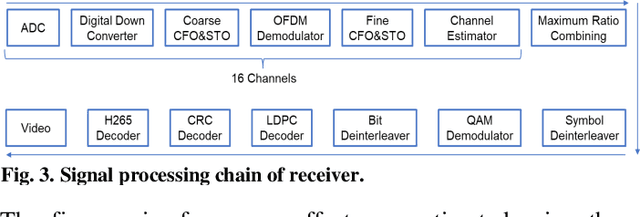
Wireless capsule endoscopy (WCE) has been widely adopted as complementary to traditional wired gastroendoscopy, especially for small bowel diseases which are beyond the latter's reach. However, both the video resolution and frame rates are limited in current WCE solutions due to the limited wireless data rate. The reasons behind this are that the electromagnetic (EM), radio frequency (RF) based communication scheme used by WCE has strict limits on useable bandwidth and power, and the high attenuation in the human body compared to air. Ultrasound communication could be a potential alternative solution as it has access to much higher bandwidths and transmitted power with much lower attenuation. In this paper, we propose an ultrasound communication scheme specially designed for high data rate through tissue data transmission and validate this communication scheme by successfully transmitting ultra-high-definition (UHD) video (3840*2160 pixels at 60 FPS) through 5 cm of pork belly. Over 8.3 Mbps error free payload data rate was achieved with the proposed communication scheme and our custom-built field programmable gate array (FPGA) based test platform.
Calibrating Data Mismatches in Deep Learning-Based Quantitative Ultrasound Using Setting Transfer Functions
Oct 04, 2022
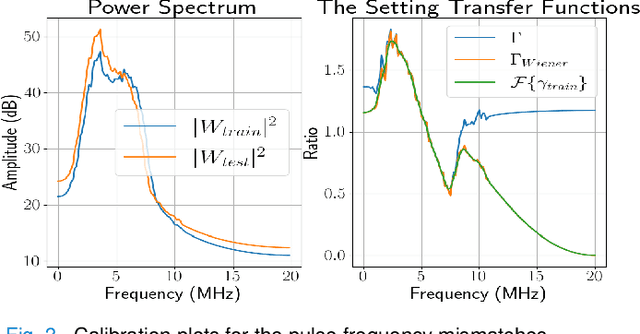
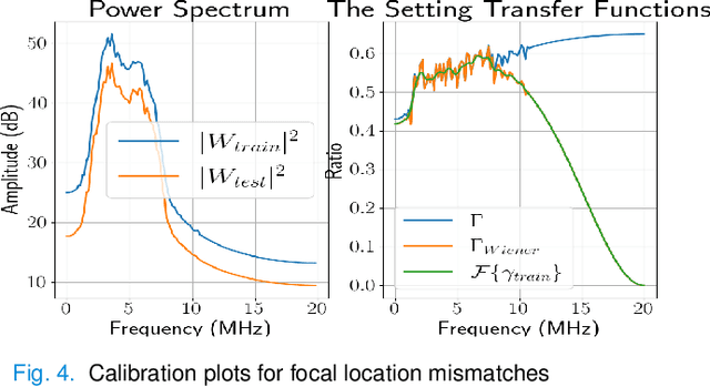
Deep learning (DL) can fail when there are data mismatches between training and testing data. Due to its operator-dependent nature, acquisition-related data mismatches, caused by different scanner settings, can occur in ultrasound imaging. Therefore, mitigating effects of such data mismatches is essential for wider clinical adoption of DL powered ultrasound imaging. To mitigate the effects, ideally we need to collect a large training set at each scanner setting. However, acquiring such training sets is expensive. Another approach could be training on a subset of imaging settings, which makes the data generation less expensive. However, there will still be generalization issues. As an alternative approach that is inexpensive and generalizable, we propose to collect a large training set at a single setting and a small calibration set at each scanner setting. Then, the calibration set will be used to calibrate data mismatches by using a signals and systems perspective. We tested the proposed solution to classify two phantoms. To investigate generalizability of the proposed solution, we calibrated three types of data mismatches: pulse frequency, focus and output power mismatches. To calibrate the setting mismatches, we calculated the setting transfer functions. The CNN trained with no calibration resulted in mean classification accuracies of 55.3%, 64.4% and 70.3% for pulse frequency, focus and output power mismatches, respectively. By using the setting transfer functions, which allowed a matching of the training and testing domains, we obtained mean accuracies of 95.3%, 92.99% and 99.32%, respectively. Therefore, the incorporation of the setting transfer functions between scanner settings can provide an economical means of generalizing a DL model for specific classification tasks where scanner settings are not fixed by the operator.