 Add to Chrome
 Add to Firefox
 Add to Edge
Compute-Optimal Network Design for Echocardiography Myocardial Segmentation and Perfusion Quantification using Neural Scaling Laws
Jun 04, 2026Myocardial perfusion quantification using contrast-enhanced ultrasound offers a bedside non-ionizing alternative to nuclear imaging modalities. However, its clinical adoption is hindered by time-consuming manual labelling. Automated segmentation has proved challenging due to a paucity of in-domain training data. Adapting strategies currently used to optimise large language models for large datasets, we apply neural scaling laws to predict network performance for myocardial segmentation. We extrapolate performance on subsets of the data to determine optimal network size on the CAMUS echocardiography dataset and a 25-patient contrast-enhanced ultrasound (CEUS) dataset. Finally, we validate the clinical utility of our models by comparing the final myocardial perfusion parameters with those obtained by a senior cardiologist. Extrapolation based on the scaling law is predictive of test loss at the full dataset size, allowing us to select two networks that obtained state-of-the-art performance on CAMUS with a 240-fold reduction in parameter count. We observe the gradient of the scaling law transfers from CAMUS to the CEUS dataset with a bias in the predicted losses. The automatically segmented masks perform equivalently to a senior cardiologist in myocardial perfusion quantification. These results establish neural scaling laws as a practical tool for data-driven compute-optimal model design for small imaging datasets.
CycleULM: A unified label-free deep learning framework for ultrasound localisation microscopy
Mar 10, 2026Super-resolution ultrasound via microbubble (MB) localisation and tracking, also known as ultrasound localisation microscopy (ULM), can resolve microvasculature beyond the acoustic diffraction limit. However, significant challenges remain in localisation performance and data acquisition and processing time. Deep learning methods for ULM have shown promise to address these challenges, however, they remain limited by in vivo label scarcity and the simulation-to-reality domain gap. We present CycleULM, the first unified label-free deep learning framework for ULM. CycleULM learns a physics-emulating translation between the real contrast-enhanced ultrasound (CEUS) data domain and a simplified MB-only domain, leveraging the power of CycleGAN without requiring paired ground truth data. With this translation, CycleULM removes dependence on high-fidelity simulators or labelled data, and makes MB localisation and tracking substantially easier. Deployed as modular plug-and-play components within existing pipelines or as an end-to-end processing framework, CycleULM delivers substantial performance gains across both in silico and in vivo datasets. Specifically, CycleULM improves image contrast (contrast-to-noise ratio) by up to 15.3 dB and sharpens CEUS resolution with a 2.5{\times} reduction in the full width at half maximum of the point spread function. CycleULM also improves MB localisation performance, with up to +40% recall, +46% precision, and a -14.0 μm mean localisation error, yielding more faithful vascular reconstructions. Importantly, CycleULM achieves real-time processing throughput at 18.3 frames per second with order-of-magnitude speed-ups (up to ~14.5{\times}). By combining label-free learning, performance enhancement, and computational efficiency, CycleULM provides a practical pathway toward robust, real-time ULM and accelerates its translation to clinical applications.
Determining the utility of ultrafast nonlinear contrast enhanced and super resolution ultrasound for imaging microcirculation in the human small intestine
May 16, 2025The regulation of intestinal blood flow is critical to gastrointestinal function. Imaging the intestinal mucosal micro-circulation in vivo has the potential to provide new insight into the gut physiology and pathophysiology. We aimed to determine whether ultrafast contrast enhanced ultrasound (CEUS) and super-resolution ultrasound localisation microscopy (SRUS/ULM) could be a useful tool for imaging the small intestine microcirculation in vivo non-invasively and for detecting changes in blood flow in the duodenum. Ultrafast CEUS and SRUS/ULM were used to image the small intestinal microcirculation in a cohort of 20 healthy volunteers (BMI<25). Participants were imaged while conscious and either having been fasted, or following ingestion of a liquid meal or water control, or under acute stress. For the first time we have performed ultrafast CEUS and ULM on the human small intestine, providing unprecedented resolution images of the intestinal microcirculation. We evaluated flow speed inside small vessels in healthy volunteers (2.78 +/- 0.05 mm/s, mean +/- SEM) and quantified changes in the perfusion of this microcirculation in response to nutrient ingestion. Perfusion of the microvasculature of the intestinal mucosa significantly increased post-prandially (36.2% +/- 12.2%, mean +/- SEM, p<0.05). The feasibility of 3D SRUS/ULM was also demonstrated. This study demonstrates the potential utility of ultrafast CEUS for assessing perfusion and detecting changes in blood flow in the duodenum. SRUS/ULM also proved a useful tool to image the microvascular blood flow in vivo non-invasively and to evaluate blood speed inside the microvasculature of the human small intestine.
Enhanced Acoustic Beamforming with Sub-Aperture Angular Multiply and Sum -- in vivo and in Human Demonstration
Jan 10, 2025


Power Doppler ultrasound is in widespread clinical use for non-invasive vascular imaging but the most common current method - Delay and Sum (DAS) beamforming - suffers from limited resolution and high side-lobes. Here we propose the Sub-Aperture Angular Multiply and Sum (SAMAS) algorithm; it combines the advantages of two recent non-linear beamformers, Frame Multiply and Sum (FMAS) which uses signal temporal coherence and the acoustic sub-aperture (ASAP) algorithm, which uses signal spatial coherence, respectively. Following in vitro experiments to optimise the algorithm, particularly the use of phase information and sub-aperture pairing, it was evaluated in vivo, first in a rabbit kidney and then in human lymph node, using ultrafast ultrasound images obtained with intravenous contrast agents. The SAMAS algorithm improved the CNR and SNR across all tests, on average raising the CNR by 11 dB and the SNR by 18 dB over DAS in vivo. This work demonstrates a promising vascular imaging method that could have widespread clinical utility.
Online 4D Ultrasound-Guided Robotic Tracking Enables 3D Ultrasound Localisation Microscopy with Large Tissue Displacements
Sep 17, 2024


Super-Resolution Ultrasound (SRUS) imaging through localising and tracking microbubbles, also known as Ultrasound Localisation Microscopy (ULM), has demonstrated significant potential for reconstructing microvasculature and flows with sub-diffraction resolution in clinical diagnostics. However, imaging organs with large tissue movements, such as those caused by respiration, presents substantial challenges. Existing methods often require breath holding to maintain accumulation accuracy, which limits data acquisition time and ULM image saturation. To improve image quality in the presence of large tissue movements, this study introduces an approach integrating high-frame-rate ultrasound with online precise robotic probe control. Tested on a microvasculature phantom with translation motions up to 20 mm, twice the aperture size of the matrix array used, our method achieved real-time tracking of the moving phantom and imaging volume rate at 85 Hz, keeping majority of the target volume in the imaging field of view. ULM images of the moving cross channels in the phantom were successfully reconstructed in post-processing, demonstrating the feasibility of super-resolution imaging under large tissue motions. This represents a significant step towards ULM imaging of organs with large motion.
Ultrafast 3-D Super Resolution Ultrasound using Row-Column Array specific Coherence-based Beamforming and Rolling Acoustic Sub-aperture Processing: In Vitro, In Vivo and Clinical Study
Nov 15, 2023


The row-column addressed array is an emerging probe for ultrafast 3-D ultrasound imaging. It achieves this with far fewer independent electronic channels and a wider field of view than traditional 2-D matrix arrays, of the same channel count, making it a good candidate for clinical translation. However, the image quality of row-column arrays is generally poor, particularly when investigating tissue. Ultrasound localisation microscopy allows for the production of super-resolution images even when the initial image resolution is not high. Unfortunately, the row-column probe can suffer from imaging artefacts that can degrade the quality of super-resolution images as `secondary' lobes from bright microbubbles can be mistaken as microbubble events, particularly when operated using plane wave imaging. These false events move through the image in a physiologically realistic way so can be challenging to remove via tracking, leading to the production of 'false vessels'. Here, a new type of rolling window image reconstruction procedure was developed, which integrated a row-column array-specific coherence-based beamforming technique with acoustic sub-aperture processing for the purposes of reducing `secondary' lobe artefacts, noise and increasing the effective frame rate. Using an {\it{in vitro}} cross tube, it was found that the procedure reduced the percentage of `false' locations from $\sim$26\% to $\sim$15\% compared to traditional orthogonal plane wave compounding. Additionally, it was found that the noise could be reduced by $\sim$7 dB and that the effective frame rate could be increased to over 4000 fps. Subsequently, {\it{in vivo}} ultrasound localisation microscopy was used to produce images non-invasively of a rabbit kidney and a human thyroid.
On the Use of Singular Value Decomposition as a Clutter Filter for Ultrasound Flow Imaging
Apr 25, 2023


Filtering based on Singular Value Decomposition (SVD) provides substantial separation of clutter, flow and noise in high frame rate ultrasound flow imaging. The use of SVD as a clutter filter has greatly improved techniques such as vector flow imaging, functional ultrasound and super-resolution ultrasound localization microscopy. The removal of clutter and noise relies on the assumption that tissue, flow and noise are each represented by different subsets of singular values, so that their signals are uncorrelated and lay on orthogonal sub-spaces. This assumption fails in the presence of tissue motion, for near-wall or microvascular flow, and can be influenced by an incorrect choice of singular value thresholds. Consequently, separation of flow, clutter and noise is imperfect, which can lead to image artefacts not present in the original data. Temporal and spatial fluctuation in intensity are the commonest artefacts, which vary in appearance and strengths. Ghosting and splitting artefacts are observed in the microvasculature where the flow signal is sparsely distributed. Singular value threshold selection, tissue motion, frame rate, flow signal amplitude and acquisition length affect the prevalence of these artefacts. Understanding what causes artefacts due to SVD clutter and noise removal is necessary for their interpretation.
Transthoracic super-resolution ultrasound localisation microscopy of myocardial vasculature in patients
Mar 28, 2023Micro-vascular flow in the myocardium is of significant importance clinically but remains poorly understood. Up to 25% of patients with symptoms of coronary heart diseases have no obstructive coronary arteries and have suspected microvascular diseases. However, such microvasculature is difficult to image in vivo with existing modalities due to the lack of resolution and sensitivity. Here, we demonstrate the feasibility of transthoracic super-resolution ultrasound localisation microscopy (SRUS/ULM) of myocardial microvasculature and hemodynamics in a large animal model and in patients, using a cardiac phased array probe with a customised data acquisition and processing pipeline. A multi-level motion correction strategy was proposed. A tracking framework incorporating multiple features and automatic parameter initialisations was developed to reconstruct microcirculation. In two patients with impaired myocardial function, we have generated SRUS images of myocardial vascular structure and flow with a resolution that is beyond the wave-diffraction limit (half a wavelength), using data acquired within a breath hold. Myocardial SRUS/ULM has potential to improve the understanding of myocardial microcirculation and the management of patients with cardiac microvascular diseases.
3D Super-Resolution Ultrasound with Adaptive Weight-Based Beamforming
Aug 25, 2022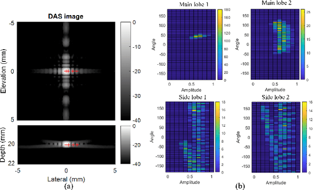
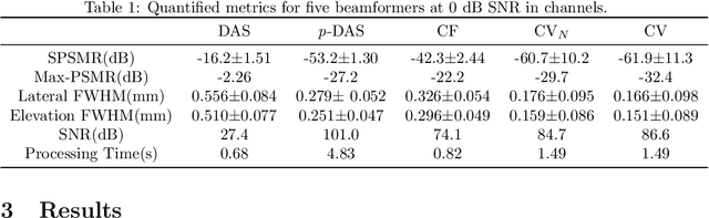
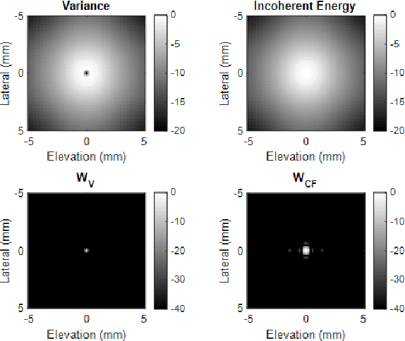
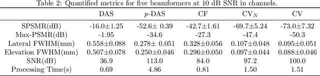
Super-resolution ultrasound (SRUS) imaging through localising and tracking sparse microbubbles has been shown to reveal microvascular structure and flow beyond the wave diffraction limit. Most SRUS studies use standard delay and sum (DAS) beamforming, where large main lobe and significant side lobes make separation and localisation of densely distributed bubbles challenging, particularly in 3D due to the typically small aperture of matrix array probes. This study aims to improve 3D SRUS by implementing a low-cost 3D coherence beamformer based on channel signal variance, as well as two other adaptive weight-based coherence beamformers: nonlinear beamforming with p-th root compression and coherence factor. The 3D coherence beamformers, together with DAS, are compared in computer simulation, on a microflow phantom, and in vivo. Simulation results demonstrate that the adaptive weight-based beamformers can significantly narrow the main lobe and suppress the side lobes for modest computational cost. Significantly improved 3D SR images of microflow phantom and a rabbit kidney are obtained through the adaptive weight-based beamformers. The proposed variance-based beamformer performs best in simulations and experiments.
Fast and selective super-resolution ultrasound in vivo with sono-switchable nanodroplets
Mar 08, 2022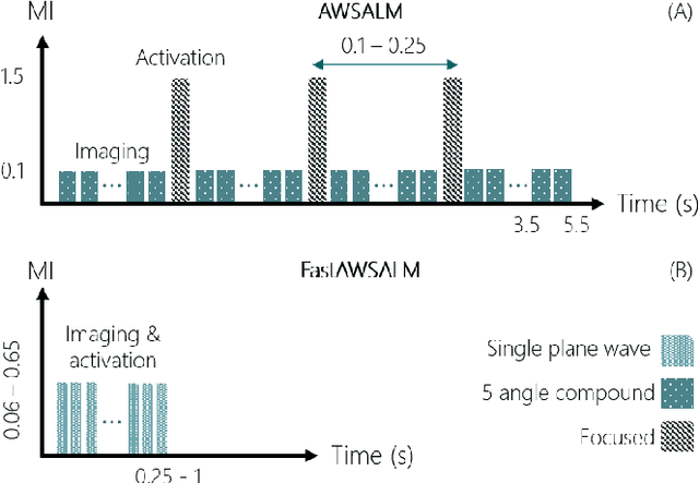
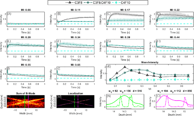
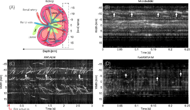
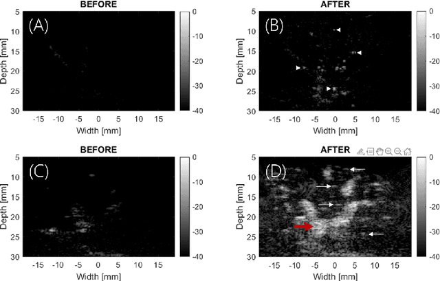
Perfusion by the microcirculation is key to the development, maintenance and pathology of tissue. Its measurement with high spatiotemporal resolution is consequently valuable but remains a challenge in deep tissue. Ultrasound Localization Microscopy (ULM) provides very high spatiotemporal resolution but the use of microbubbles requires low contrast agent concentrations, a long acquisition time, and gives little control over the spatial and temporal distribution of the bubbles. The present study is the first to demonstrate Acoustic Wave Sparsely-Activated Localization Microscopy (AWSALM) and fast-AWSALM for in vivo super-resolution ultrasound imaging, offering contrast on demand and vascular selectivity. Three different formulations of sono-switchable contrast agents were tested. We demonstrate their use with ultrasound mechanical indices well within recommended safety limits to enable fast on-demand sparse switching at very high agent concentrations. We produce super-localization maps of the rabbit renal vasculature with acquisition times between 5.5 s and 0.25 s, and an 4-fold improvement in spatial resolution. We present the unique selectivity of AWSALM in visualizing specific vascular branches and downstream microvasculature, and we show super-localized kidney structures in systole and diastole with fast-AWSALM. In conclusion we demonstrate the feasibility of fast and selective measurement of microvascular dynamics in vivo with subwavelength resolution using ultrasound and sono-switchable nanodroplets.