 Add to Chrome
 Add to Firefox
 Add to Edge
Compute-Optimal Network Design for Echocardiography Myocardial Segmentation and Perfusion Quantification using Neural Scaling Laws
Jun 04, 2026Myocardial perfusion quantification using contrast-enhanced ultrasound offers a bedside non-ionizing alternative to nuclear imaging modalities. However, its clinical adoption is hindered by time-consuming manual labelling. Automated segmentation has proved challenging due to a paucity of in-domain training data. Adapting strategies currently used to optimise large language models for large datasets, we apply neural scaling laws to predict network performance for myocardial segmentation. We extrapolate performance on subsets of the data to determine optimal network size on the CAMUS echocardiography dataset and a 25-patient contrast-enhanced ultrasound (CEUS) dataset. Finally, we validate the clinical utility of our models by comparing the final myocardial perfusion parameters with those obtained by a senior cardiologist. Extrapolation based on the scaling law is predictive of test loss at the full dataset size, allowing us to select two networks that obtained state-of-the-art performance on CAMUS with a 240-fold reduction in parameter count. We observe the gradient of the scaling law transfers from CAMUS to the CEUS dataset with a bias in the predicted losses. The automatically segmented masks perform equivalently to a senior cardiologist in myocardial perfusion quantification. These results establish neural scaling laws as a practical tool for data-driven compute-optimal model design for small imaging datasets.
Transthoracic super-resolution ultrasound localisation microscopy of myocardial vasculature in patients
Mar 28, 2023Micro-vascular flow in the myocardium is of significant importance clinically but remains poorly understood. Up to 25% of patients with symptoms of coronary heart diseases have no obstructive coronary arteries and have suspected microvascular diseases. However, such microvasculature is difficult to image in vivo with existing modalities due to the lack of resolution and sensitivity. Here, we demonstrate the feasibility of transthoracic super-resolution ultrasound localisation microscopy (SRUS/ULM) of myocardial microvasculature and hemodynamics in a large animal model and in patients, using a cardiac phased array probe with a customised data acquisition and processing pipeline. A multi-level motion correction strategy was proposed. A tracking framework incorporating multiple features and automatic parameter initialisations was developed to reconstruct microcirculation. In two patients with impaired myocardial function, we have generated SRUS images of myocardial vascular structure and flow with a resolution that is beyond the wave-diffraction limit (half a wavelength), using data acquired within a breath hold. Myocardial SRUS/ULM has potential to improve the understanding of myocardial microcirculation and the management of patients with cardiac microvascular diseases.
Fully Automatic Myocardial Segmentation of Contrast Echocardiography Sequence Using Random Forests Guided by Shape Model
Jun 19, 2018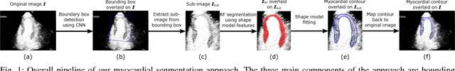
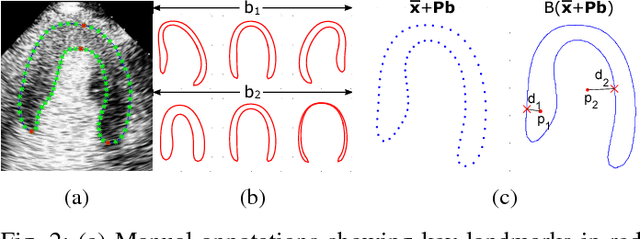
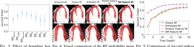
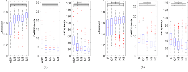
Myocardial contrast echocardiography (MCE) is an imaging technique that assesses left ventricle function and myocardial perfusion for the detection of coronary artery diseases. Automatic MCE perfusion quantification is challenging and requires accurate segmentation of the myocardium from noisy and time-varying images. Random forests (RF) have been successfully applied to many medical image segmentation tasks. However, the pixel-wise RF classifier ignores contextual relationships between label outputs of individual pixels. RF which only utilizes local appearance features is also susceptible to data suffering from large intensity variations. In this paper, we demonstrate how to overcome the above limitations of classic RF by presenting a fully automatic segmentation pipeline for myocardial segmentation in full-cycle 2D MCE data. Specifically, a statistical shape model is used to provide shape prior information that guide the RF segmentation in two ways. First, a novel shape model (SM) feature is incorporated into the RF framework to generate a more accurate RF probability map. Second, the shape model is fitted to the RF probability map to refine and constrain the final segmentation to plausible myocardial shapes. We further improve the performance by introducing a bounding box detection algorithm as a preprocessing step in the segmentation pipeline. Our approach on 2D image is further extended to 2D+t sequence which ensures temporal consistency in the resultant sequence segmentations. When evaluated on clinical MCE data, our proposed method achieves notable improvement in segmentation accuracy and outperforms other state-of-the-art methods including the classic RF and its variants, active shape model and image registration.
Myocardial Segmentation of Contrast Echocardiograms Using Random Forests Guided by Shape Model
Jun 19, 2018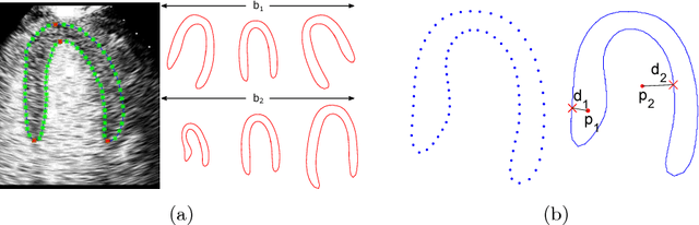
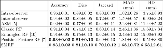
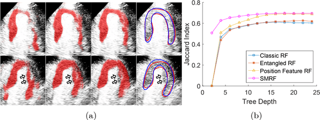
Myocardial Contrast Echocardiography (MCE) with micro-bubble contrast agent enables myocardial perfusion quantification which is invaluable for the early detection of coronary artery diseases. In this paper, we proposed a new segmentation method called Shape Model guided Random Forests (SMRF) for the analysis of MCE data. The proposed method utilizes a statistical shape model of the myocardium to guide the Random Forest (RF) segmentation in two ways. First, we introduce a novel Shape Model (SM) feature which captures the global structure and shape of the myocardium to produce a more accurate RF probability map. Second, the shape model is fitted to the RF probability map to further refine and constrain the final segmentation to plausible myocardial shapes. Evaluated on clinical MCE images from 15 patients, our method obtained promising results (Dice=0.81, Jaccard=0.70, MAD=1.68 mm, HD=6.53 mm) and showed a notable improvement in segmentation accuracy over the classic RF and its variants.