 Add to Chrome
 Add to Firefox
 Add to Edge
CytoSyn: a Foundation Diffusion Model for Histopathology -- Tech Report
Mar 18, 2026Computational pathology has made significant progress in recent years, fueling advances in both fundamental disease understanding and clinically ready tools. This evolution is driven by the availability of large amounts of digitized slides and specialized deep learning methods and models. Multiple self-supervised foundation feature extractors have been developed, enabling downstream predictive applications from cell segmentation to tumor sub-typing and survival analysis. In contrast, generative foundation models designed specifically for histopathology remain scarce. Such models could address tasks that are beyond the capabilities of feature extractors, such as virtual staining. In this paper, we introduce CytoSyn, a state-of-the-art foundation latent diffusion model that enables the guided generation of highly realistic and diverse histopathology H&E-stained images, as shown in an extensive benchmark. We explored methodological improvements, training set scaling, sampling strategies and slide-level overfitting, culminating in the improved CytoSyn-v2, and compared our work to PixCell, a state-of-the-art model, in an in-depth manner. This comparison highlighted the strong sensitivity of both diffusion models and performance metrics to preprocessing-specific details such as JPEG compression. Our model has been trained on a dataset obtained from more than 10,000 TCGA diagnostic whole-slide images of 32 different cancer types. Despite being trained only on oncology slides, it maintains state-of-the-art performance generating inflammatory bowel disease images. To support the research community, we publicly release CytoSyn's weights, its training and validation datasets, and a sample of synthetic images in this repository: https://huggingface.co/Owkin-Bioptimus/CytoSyn.
Using StyleGAN for Visual Interpretability of Deep Learning Models on Medical Images
Jan 19, 2021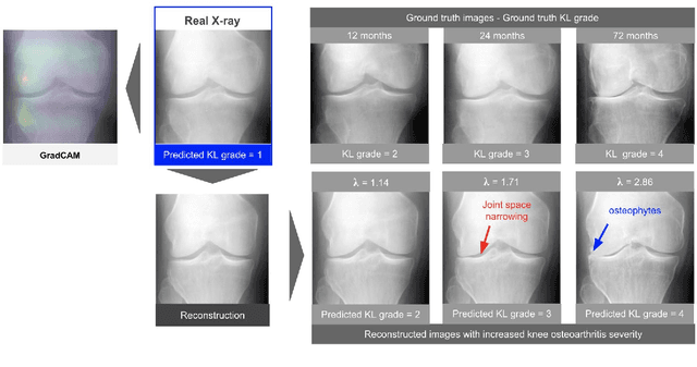
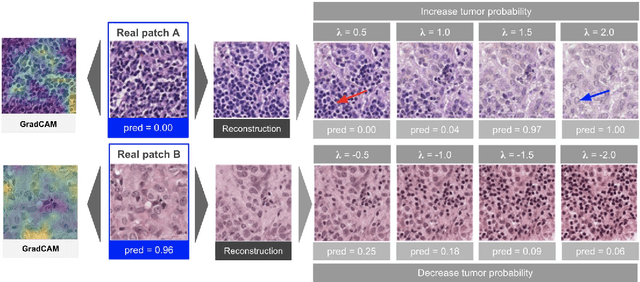
As AI-based medical devices are becoming more common in imaging fields like radiology and histology, interpretability of the underlying predictive models is crucial to expand their use in clinical practice. Existing heatmap-based interpretability methods such as GradCAM only highlight the location of predictive features but do not explain how they contribute to the prediction. In this paper, we propose a new interpretability method that can be used to understand the predictions of any black-box model on images, by showing how the input image would be modified in order to produce different predictions. A StyleGAN is trained on medical images to provide a mapping between latent vectors and images. Our method identifies the optimal direction in the latent space to create a change in the model prediction. By shifting the latent representation of an input image along this direction, we can produce a series of new synthetic images with changed predictions. We validate our approach on histology and radiology images, and demonstrate its ability to provide meaningful explanations that are more informative than GradCAM heatmaps. Our method reveals the patterns learned by the model, which allows clinicians to build trust in the model's predictions, discover new biomarkers and eventually reveal potential biases.
Statistical learning of spatiotemporal patterns from longitudinal manifold-valued networks
Sep 25, 2017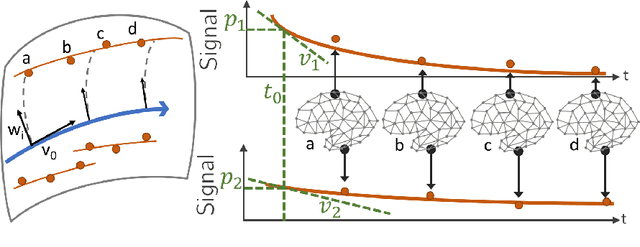
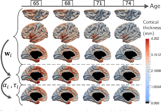
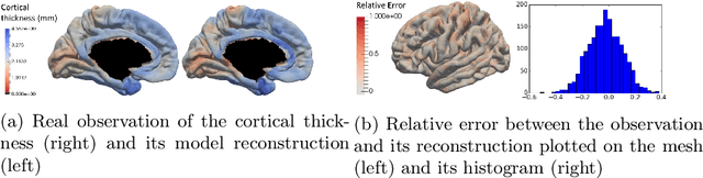
We introduce a mixed-effects model to learn spatiotempo-ral patterns on a network by considering longitudinal measures distributed on a fixed graph. The data come from repeated observations of subjects at different time points which take the form of measurement maps distributed on a graph such as an image or a mesh. The model learns a typical group-average trajectory characterizing the propagation of measurement changes across the graph nodes. The subject-specific trajectories are defined via spatial and temporal transformations of the group-average scenario, thus estimating the variability of spatiotemporal patterns within the group. To estimate population and individual model parameters, we adapted a stochastic version of the Expectation-Maximization algorithm, the MCMC-SAEM. The model is used to describe the propagation of cortical atrophy during the course of Alzheimer's Disease. Model parameters show the variability of this average pattern of atrophy in terms of trajectories across brain regions, age at disease onset and pace of propagation. We show that the personalization of this model yields accurate prediction of maps of cortical thickness in patients.