 Add to Chrome
 Add to Firefox
 Add to Edge
Deep Learning-Based Automatic Diagnosis System for Developmental Dysplasia of the Hip
Sep 07, 2022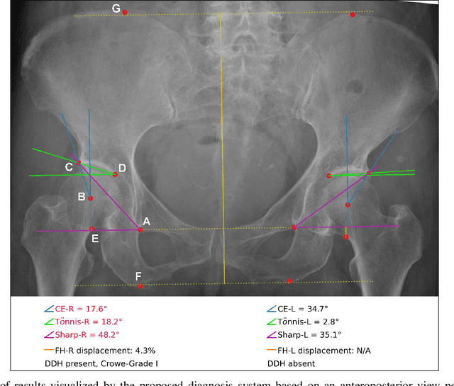
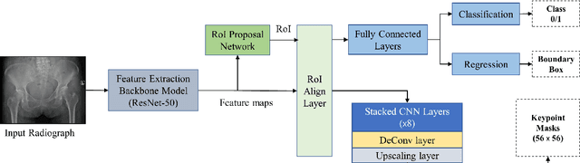
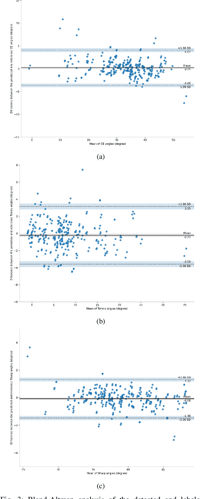
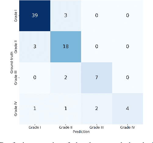
As the first-line diagnostic imaging modality, radiography plays an essential role in the early detection of developmental dysplasia of the hip (DDH). Clinically, the diagnosis of DDH relies on manual measurements and subjective evaluation of different anatomical features from pelvic radiographs. This process is inefficient and error-prone and requires years of clinical experience. In this study, we propose a deep learning-based system that automatically detects 14 keypoints from a radiograph, measures three anatomical angles (center-edge, T\"onnis, and Sharp angles), and classifies DDH hips as grades I-IV based on the Crowe criteria. Moreover, a novel data-driven scoring system is proposed to quantitatively integrate the information from the three angles for DDH diagnosis. The proposed keypoint detection model achieved a mean (95% confidence interval [CI]) average precision of 0.807 (0.804-0.810). The mean (95% CI) intraclass correlation coefficients between the center-edge, Tonnis, and Sharp angles measured by the proposed model and the ground-truth were 0.957 (0.952-0.962), 0.947 (0.941-0.953), and 0.953 (0.947-0.960), respectively, which were significantly higher than those of experienced orthopedic surgeons (p<0.0001). In addition, the mean (95% CI) test diagnostic agreement (Cohen's kappa) obtained using the proposed scoring system was 0.84 (0.83-0.85), which was significantly higher than those obtained from diagnostic criteria for individual angle (0.76 [0.75-0.77]) and orthopedists (0.71 [0.63-0.79]). To the best of our knowledge, this is the first study for objective DDH diagnosis by leveraging deep learning keypoint detection and integrating different anatomical measurements, which can provide reliable and explainable support for clinical decision-making.
Machine learning of percolation models using graph convolutional neural networks
Jul 07, 2022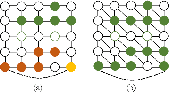
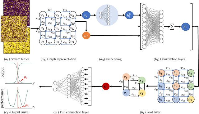
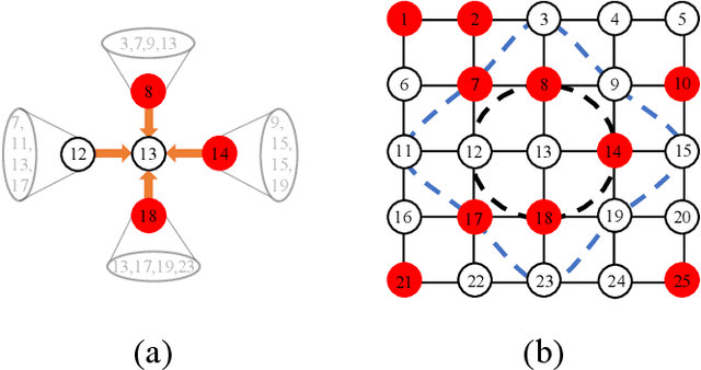
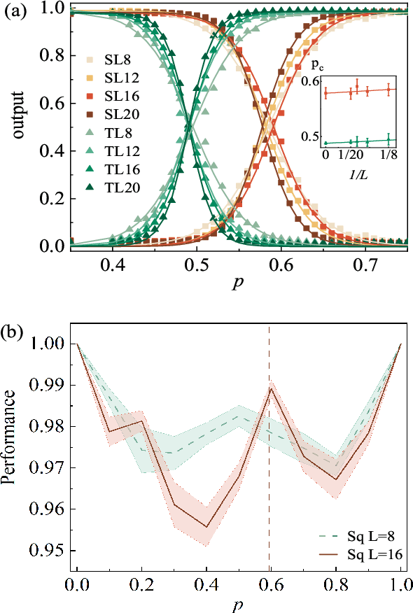
Percolation is an important topic in climate, physics, materials science, epidemiology, finance, and so on. Prediction of percolation thresholds with machine learning methods remains challenging. In this paper, we build a powerful graph convolutional neural network to study the percolation in both supervised and unsupervised ways. From a supervised learning perspective, the graph convolutional neural network simultaneously and correctly trains data of different lattice types, such as the square and triangular lattices. For the unsupervised perspective, combining the graph convolutional neural network and the confusion method, the percolation threshold can be obtained by the "W" shaped performance. The finding of this work opens up the possibility of building a more general framework that can probe the percolation-related phenomenon.
Deep Learning-based End-to-end Diagnosis System for Avascular Necrosis of Femoral Head
Feb 12, 2020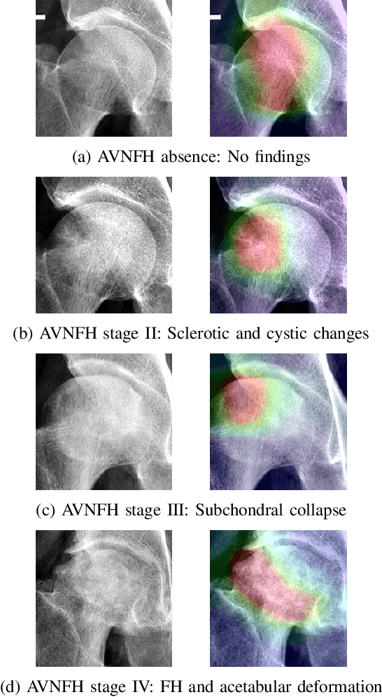
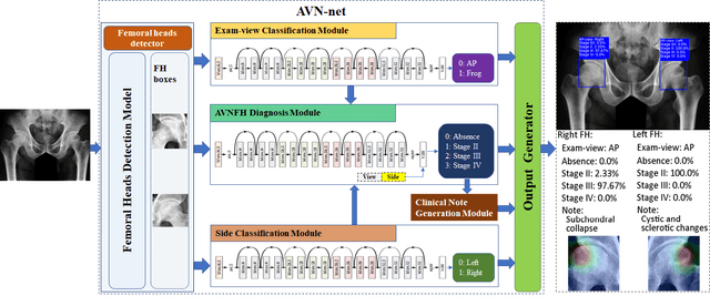
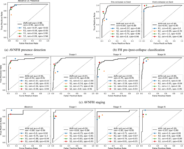
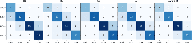
As the first diagnostic imaging modality of avascular necrosis of the femoral head (AVNFH), accurately staging AVNFH from a plain radiograph is critical and challenging for orthopedists. Thus, we propose a deep learning-based AVNFH diagnosis system (AVN-net). The proposed AVN-net reads plain radiographs of the pelvis, conducts diagnosis, and visualizes results automatically. Deep convolutional neural networks are trained to provide an end-to-end diagnosis solution, covering femoral head detection, exam-view/sides identification, AVNFH diagnosis, and key clinical note generation subtasks. AVN-net is able to obtain state-of-the-art testing AUC of 0.95 (95% CI: 0.92-0.98) in AVNFH detection and significantly greater F1 scores (p<0.01) than less-to-moderately experienced orthopedists in all diagnostic tests. Furthermore, two real-world pilot studies were conducted for diagnosis support and education assistance, respectively, to assess the utility of AVN-net. The experimental results are promising. With the AVN-net diagnosis as a reference, the diagnostic accuracy and consistency of all orthopedists considerably improved while requiring only 1/4 of the time. Students self-studying the AVNFH diagnosis using AVN-net can learn better and faster than the control group. To the best of our knowledge, this study is the first research on the prospective use of a deep learning-based diagnosis system for AVNFH by conducting two pilot studies representing real-world application scenarios. We have demonstrated that the proposed AVN-net achieves expert-level AVNFH diagnosis performance, provides efficient support in clinical decision-making, and effectively passes clinical experience to students.
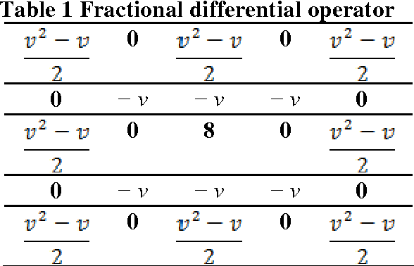
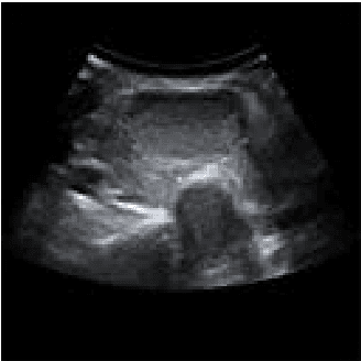
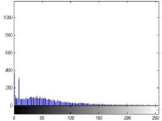
Segmentation of ultrasound images of thyroid nodule for assisting fine needle aspiration cytology
Nov 03, 2012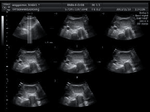
The incidence of thyroid nodule is very high and generally increases with the age. Thyroid nodule may presage the emergence of thyroid cancer. The thyroid nodule can be completely cured if detected early. Fine needle aspiration cytology is a recognized early diagnosis method of thyroid nodule. There are still some limitations in the fine needle aspiration cytology, and the ultrasound diagnosis of thyroid nodule has become the first choice for auxiliary examination of thyroid nodular disease. If we could combine medical imaging technology and fine needle aspiration cytology, the diagnostic rate of thyroid nodule would be improved significantly. The properties of ultrasound will degrade the image quality, which makes it difficult to recognize the edges for physicians. Image segmentation technique based on graph theory has become a research hotspot at present. Normalized cut (Ncut) is a representative one, which is suitable for segmentation of feature parts of medical image. However, how to solve the normalized cut has become a problem, which needs large memory capacity and heavy calculation of weight matrix. It always generates over segmentation or less segmentation which leads to inaccurate in the segmentation. The speckle noise in B ultrasound image of thyroid tumor makes the quality of the image deteriorate. In the light of this characteristic, we combine the anisotropic diffusion model with the normalized cut in this paper. After the enhancement of anisotropic diffusion model, it removes the noise in the B ultrasound image while preserves the important edges and local details. This reduces the amount of computation in constructing the weight matrix of the improved normalized cut and improves the accuracy of the final segmentation results. The feasibility of the method is proved by the experimental results.