 Add to Chrome
 Add to Firefox
 Add to Edge
Dense Temporal Contrast Synthesis via Conditioned Latent Transport
Jul 31, 2026
Dynamic contrast-enhanced magnetic resonance imaging (DCE-MRI) is essential for breast cancer management, but reliance on gadolinium-based contrast agents (GBCAs) restricts use in contraindicated populations, prolongs scan protocols, and presents environmental toxicity concerns. Contrast synthesis offers a non-invasive alternative; however, existing approaches struggle to balance spatial realism with temporal continuity, suffer from slow iterative sampling, underutilize structural priors, and lack clinical validation. We propose a novel conditioned latent transport framework that predicts contrast enhancement in a single forward pass. By anchoring the latent trajectory to the pre-contrast anatomy and applying continuous time conditioning, the model synthesizes patient-specific contrast evolution at any acquisition time. The proposed approach outperforms baseline and the state-of-the-art models across spatial, perceptual, temporal, and distributional metrics. Evaluated on an independent external cohort, the method demonstrates robustness to domain shifts induced by scanner noise as well as differing acquisition protocol. Furthermore, our synthetic contrast enhancement significantly improved downstream tumor segmentation performance, yielding a 22.4% relative increase in Dice coefficient (0.60 vs. 0.49 baseline pre-contrast, p < 0.01), reducing boundary segmentation error by over 39%, while outperforming all other generative model baselines. Finally, a reader study involving four breast radiologists evaluated the image quality, kinetic fidelity, and diagnostic viability of our synthesized sequences across 40 randomly selected cases. The results demonstrated that in 70% of cases, synthesized images provided sufficient clinical information to support the same management decisions as real DCE-MRI, suggesting a path toward safer and faster contrast-free or contrast-reduced imaging workflows.

Graph Representation Learning of Longitudinal Medical Imaging Trajectories for Treatment Response Prediction
Jul 06, 2026In patients with breast cancer, pathological complete response (pCR) has been established as a clinically meaningful surrogate marker for long-term outcomes. While commonly treated with neoadjuvant chemotherapy (NACT), effective treatment decision-making remains challenging, as therapeutic response can vary substantially across patients, calling for predictive models capable of accurately estimating individualized treatment response. To address this, we propose an imaging-based 3D spatio-temporal framework for treatment response prediction that integrates a state-of-the-art graph neural network with relational modeling of temporal interactions across timepoints alongside three novel complementary self-supervised treatment trajectory representation learning objectives. Experiments across a cohort of 585 patients from the public ISPY-2 dataset demonstrate that our method substantially outperforms both vision and self-supervised learning baselines across several classification metrics. Alongside establishing a breast cancer pCR prediction benchmark, we include a principled ablation of our method and further introduce and empirically assess the impact of the available number of DCE-MRI timepoints per patient trajectory and the inclusion of inter-scan time-differences. Overall, our study substantiates the utility of clinically meaningful longitudinal medical imagaging modeling for predicting NACT-induced pCR. We will publicly share our code repository and a user-friendly PyPI library for dataset curation upon publication, effectively promoting reproducible open-source research.
Entropy Minimization without Model Collapse: Mitigating Prediction Bias in Medical Imaging
Jun 01, 2026Entropy minimization (EM) is the dominant objective for test-time adaptation, yet its failure mode, model collapse, remains poorly understood. In this work, we show that distribution shifts can cause feature clusters corresponding to distinct classes in the model's representation space to merge, while the decision boundary remains fixed. This induces a systematic skew in the predicted class distribution, referred to as prediction bias. Prediction bias refers to a shift in the predicted class distribution, with some classes overrepresented and others suppressed. We show that entropy minimization amplifies this prediction bias by tightening the existing clusters, reinforcing the incorrect groupings until all predictions collapse to a trivial solution. Next, to demonstrate the significance of prediction bias and mitigate it, we further propose Distribution Shift Bias Reduction (DSBR), a bias-correcting objective that specifically targets this failure mode by equalizing the contribution of each predicted class to the unsupervised entropy minimization loss. To study this failure mode, we design suitable adaptation settings using four medical-imaging datasets and additionally evaluate on ImageNet-C. We find that DSBR consistently stabilizes test-time adaptation, prevents model collapse, and matches or outperforms state-of-the-art methods. Moreover, DSBR operates solely at test-time.
The Mean is the Mirage: Entropy-Adaptive Model Merging under Heterogeneous Domain Shifts in Medical Imaging
Feb 24, 2026Model merging under unseen test-time distribution shifts often renders naive strategies, such as mean averaging unreliable. This challenge is especially acute in medical imaging, where models are fine-tuned locally at clinics on private data, producing domain-specific models that differ by scanner, protocol, and population. When deployed at an unseen clinical site, test cases arrive in unlabeled, non-i.i.d. batches, and the model must adapt immediately without labels. In this work, we introduce an entropy-adaptive, fully online model-merging method that yields a batch-specific merged model via only forward passes, effectively leveraging target information. We further demonstrate why mean merging is prone to failure and misaligned under heterogeneous domain shifts. Next, we mitigate encoder classifier mismatch by decoupling the encoder and classification head, merging with separate merging coefficients. We extensively evaluate our method with state-of-the-art baselines using two backbones across nine medical and natural-domain generalization image classification datasets, showing consistent gains across standard evaluation and challenging scenarios. These performance gains are achieved while retaining single-model inference at test-time, thereby demonstrating the effectiveness of our method.
TomoGraphView: 3D Medical Image Classification with Omnidirectional Slice Representations and Graph Neural Networks
Nov 12, 2025The growing number of medical tomography examinations has necessitated the development of automated methods capable of extracting comprehensive imaging features to facilitate downstream tasks such as tumor characterization, while assisting physicians in managing their growing workload. However, 3D medical image classification remains a challenging task due to the complex spatial relationships and long-range dependencies inherent in volumetric data. Training models from scratch suffers from low data regimes, and the absence of 3D large-scale multimodal datasets has limited the development of 3D medical imaging foundation models. Recent studies, however, have highlighted the potential of 2D vision foundation models, originally trained on natural images, as powerful feature extractors for medical image analysis. Despite these advances, existing approaches that apply 2D models to 3D volumes via slice-based decomposition remain suboptimal. Conventional volume slicing strategies, which rely on canonical planes such as axial, sagittal, or coronal, may inadequately capture the spatial extent of target structures when these are misaligned with standardized viewing planes. Furthermore, existing slice-wise aggregation strategies rarely account for preserving the volumetric structure, resulting in a loss of spatial coherence across slices. To overcome these limitations, we propose TomoGraphView, a novel framework that integrates omnidirectional volume slicing with spherical graph-based feature aggregation. We publicly share our accessible code base at http://github.com/compai-lab/2025-MedIA-kiechle and provide a user-friendly library for omnidirectional volume slicing at https://pypi.org/project/OmniSlicer.
Simulating Dynamic Tumor Contrast Enhancement in Breast MRI using Conditional Generative Adversarial Networks
Sep 27, 2024This paper presents a method for virtual contrast enhancement in breast MRI, offering a promising non-invasive alternative to traditional contrast agent-based DCE-MRI acquisition. Using a conditional generative adversarial network, we predict DCE-MRI images, including jointly-generated sequences of multiple corresponding DCE-MRI timepoints, from non-contrast-enhanced MRIs, enabling tumor localization and characterization without the associated health risks. Furthermore, we qualitatively and quantitatively evaluate the synthetic DCE-MRI images, proposing a multi-metric Scaled Aggregate Measure (SAMe), assessing their utility in a tumor segmentation downstream task, and conclude with an analysis of the temporal patterns in multi-sequence DCE-MRI generation. Our approach demonstrates promising results in generating realistic and useful DCE-MRI sequences, highlighting the potential of virtual contrast enhancement for improving breast cancer diagnosis and treatment, particularly for patients where contrast agent administration is contraindicated.
Graph Neural Networks: A suitable Alternative to MLPs in Latent 3D Medical Image Classification?
Jul 24, 2024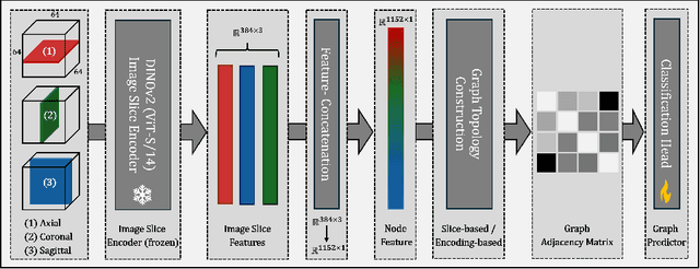
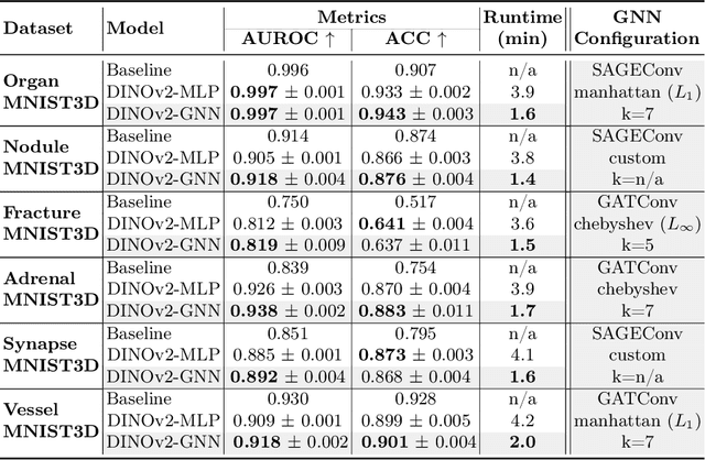
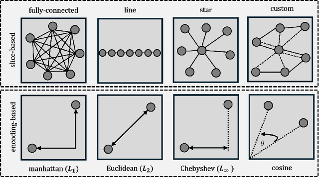
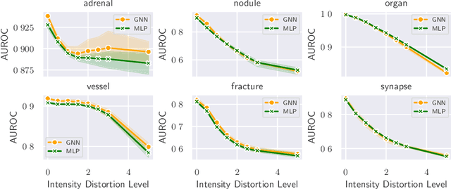
Recent studies have underscored the capabilities of natural imaging foundation models to serve as powerful feature extractors, even in a zero-shot setting for medical imaging data. Most commonly, a shallow multi-layer perceptron (MLP) is appended to the feature extractor to facilitate end-to-end learning and downstream prediction tasks such as classification, thus representing the de facto standard. However, as graph neural networks (GNNs) have become a practicable choice for various tasks in medical research in the recent past, we direct attention to the question of how effective GNNs are compared to MLP prediction heads for the task of 3D medical image classification, proposing them as a potential alternative. In our experiments, we devise a subject-level graph for each volumetric dataset instance. Therein latent representations of all slices in the volume, encoded through a DINOv2 pretrained vision transformer (ViT), constitute the nodes and their respective node features. We use public datasets to compare the classification heads numerically and evaluate various graph construction and graph convolution methods in our experiments. Our findings show enhancements of the GNN in classification performance and substantial improvements in runtime compared to an MLP prediction head. Additional robustness evaluations further validate the promising performance of the GNN, promoting them as a suitable alternative to traditional MLP classification heads. Our code is publicly available at: https://github.com/compai-lab/2024-miccai-grail-kiechle
MedEdit: Counterfactual Diffusion-based Image Editing on Brain MRI
Jul 21, 2024
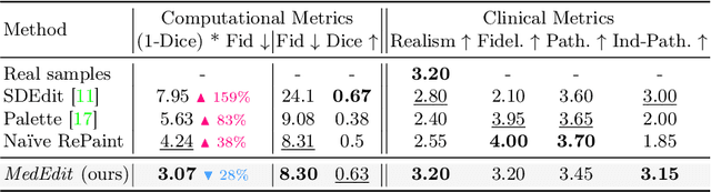
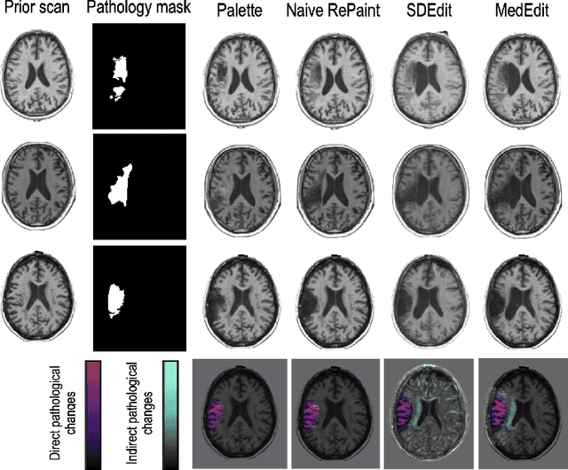
Denoising diffusion probabilistic models enable high-fidelity image synthesis and editing. In biomedicine, these models facilitate counterfactual image editing, producing pairs of images where one is edited to simulate hypothetical conditions. For example, they can model the progression of specific diseases, such as stroke lesions. However, current image editing techniques often fail to generate realistic biomedical counterfactuals, either by inadequately modeling indirect pathological effects like brain atrophy or by excessively altering the scan, which disrupts correspondence to the original images. Here, we propose MedEdit, a conditional diffusion model for medical image editing. MedEdit induces pathology in specific areas while balancing the modeling of disease effects and preserving the integrity of the original scan. We evaluated MedEdit on the Atlas v2.0 stroke dataset using Frechet Inception Distance and Dice scores, outperforming state-of-the-art diffusion-based methods such as Palette (by 45%) and SDEdit (by 61%). Additionally, clinical evaluations by a board-certified neuroradiologist confirmed that MedEdit generated realistic stroke scans indistinguishable from real ones. We believe this work will enable counterfactual image editing research to further advance the development of realistic and clinically useful imaging tools.
Enhancing the Utility of Privacy-Preserving Cancer Classification using Synthetic Data
Jul 17, 2024Deep learning holds immense promise for aiding radiologists in breast cancer detection. However, achieving optimal model performance is hampered by limitations in availability and sharing of data commonly associated to patient privacy concerns. Such concerns are further exacerbated, as traditional deep learning models can inadvertently leak sensitive training information. This work addresses these challenges exploring and quantifying the utility of privacy-preserving deep learning techniques, concretely, (i) differentially private stochastic gradient descent (DP-SGD) and (ii) fully synthetic training data generated by our proposed malignancy-conditioned generative adversarial network. We assess these methods via downstream malignancy classification of mammography masses using a transformer model. Our experimental results depict that synthetic data augmentation can improve privacy-utility tradeoffs in differentially private model training. Further, model pretraining on synthetic data achieves remarkable performance, which can be further increased with DP-SGD fine-tuning across all privacy guarantees. With this first in-depth exploration of privacy-preserving deep learning in breast imaging, we address current and emerging clinical privacy requirements and pave the way towards the adoption of private high-utility deep diagnostic models. Our reproducible codebase is publicly available at https://github.com/RichardObi/mammo_dp.
Progressive Growing of Patch Size: Resource-Efficient Curriculum Learning for Dense Prediction Tasks
Jul 11, 2024In this work, we introduce Progressive Growing of Patch Size, a resource-efficient implicit curriculum learning approach for dense prediction tasks. Our curriculum approach is defined by growing the patch size during model training, which gradually increases the task's difficulty. We integrated our curriculum into the nnU-Net framework and evaluated the methodology on all 10 tasks of the Medical Segmentation Decathlon. With our approach, we are able to substantially reduce runtime, computational costs, and CO2 emissions of network training compared to classical constant patch size training. In our experiments, the curriculum approach resulted in improved convergence. We are able to outperform standard nnU-Net training, which is trained with constant patch size, in terms of Dice Score on 7 out of 10 MSD tasks while only spending roughly 50% of the original training runtime. To the best of our knowledge, our Progressive Growing of Patch Size is the first successful employment of a sample-length curriculum in the form of patch size in the field of computer vision. Our code is publicly available at https://github.com/compai-lab/2024-miccai-fischer.