 Add to Chrome
 Add to Firefox
 Add to Edge
Eversion-based robots can enable safe access,steering and endoscopic imaging within the spinal subarachnoid space
Jul 21, 2026
Safe navigation within the spinal subarachnoid space is constrained by its narrow, compliant, and delicate anatomy. Conventional catheters and continuum robots rely on proximal pushing, generating friction and shear along the tissue device interface that limit distal controllability and increase the risk of neural injury. Here, we present a 2 mm diameter eversion-growing robotic platform that enables friction minimised extension and steering within the human spinal subarachnoid space, validated through computational modelling, phantom experiments, and intact human cadaver studies. The robot integrates a miniature endoscope for real time intrathecal visualisation and advances by pressure driven tip eversion, localising motion to the distal tip while minimising translational sliding of the deployed body. Phantom experiments demonstrated reductions of 65.2% in mean interaction force and 48.0% in peak interaction force compared with matched push-based insertion. Physics based modelling showed that eversion based growth redistributed tissue loading, reducing local stress concentrations and interfacial shear relative to conventional insertion. In an intact human cadaver, the system achieved 150 mm of controlled intrathecal extension with concurrent fluoroscopic and endoscopic visualisation, providing access across multiple vertebral levels from a standard lumbar entry point. Postprocedural laminectomy and durotomy revealed no observable macroscopic disruption of the dura mater or surrounding neural structures. These results provide the first mechanically characterised and multimodally validated demonstration of eversion-based robotic navigation in intact human spinal anatomy, establishing a quantitative and procedural foundation for future intrathecal interventions. Further validation in larger anatomical cohorts and under physiological conditions will be required before clinical translation.

Ultrathin, high-speed, all-optical photoacoustic endomicroscopy probe for guiding minimally invasive surgery
May 06, 2022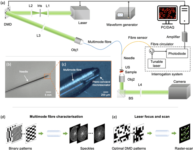
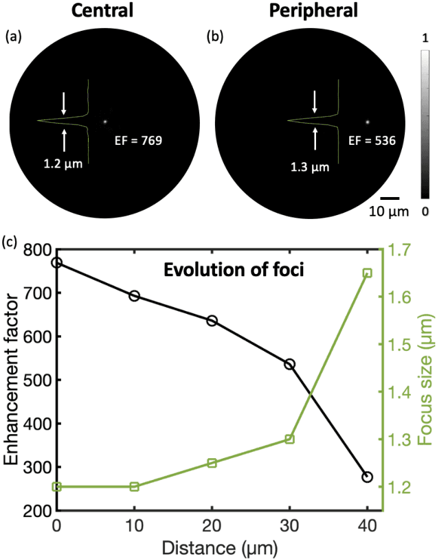
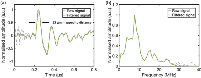
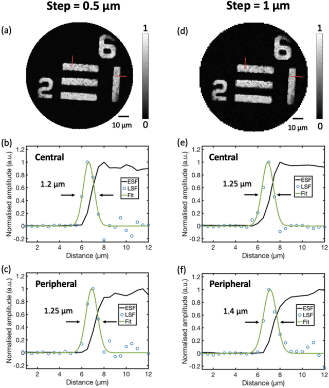
Photoacoustic (PA) endoscopy has shown significant potential for clinical diagnosis and surgical guidance. Multimode fibres (MMFs) are becoming increasing attractive for the development of miniature endoscopy probes owing to ultrathin size, low cost and diffraction-limited spatial resolution enabled by wavefront shaping. However, current MMF-based PA endomicroscopy probes are either limited by a bulky ultrasound detector or a low imaging speed which hindered their usability. In this work, we report the development of a highly miniaturised and high-speed PA endomicroscopy probe that is integrated within the cannula of a 20 gauge medical needle. This probe comprises a MMF for delivering the PA excitation light and a single-mode optical fibre with a plano-concave microresonator for ultrasound detection. Wavefront shaping with a digital micromirror device enabled rapid raster-scanning of a focused light spot at the distal end of the MMF for tissue interrogation. High-resolution PA imaging of mouse red blood cells covering an area 100 microns in diameter was achieved with the needle probe at ~3 frames per second. Mosaicing imaging was performed after fibre characterisation by translating the needle probe to enlarge the field-of-view in real-time. The developed ultrathin PA endomicroscopy probe is promising for guiding minimally invasive surgery by providing functional, molecular and microstructural information of tissue in real-time.