 Add to Chrome
 Add to Firefox
 Add to Edge
Longitudinal Risk Prediction in Mammography with Privileged History Distillation
Mar 16, 2026Breast cancer remains a leading cause of cancer-related mortality worldwide. Longitudinal mammography risk prediction models improve multi-year breast cancer risk prediction based on prior screening exams. However, in real-world clinical practice, longitudinal histories are often incomplete, irregular, or unavailable due to missed screenings, first-time examinations, heterogeneous acquisition schedules, or archival constraints. The absence of prior exams degrades the performance of longitudinal risk models and limits their practical applicability. While substantial longitudinal history is available during training, prior exams are commonly absent at test time. In this paper, we address missing history at inference time and propose a longitudinal risk prediction method that uses mammography history as privileged information during training and distills its prognostic value into a student model that only requires the current exam at inference time. The key idea is a privileged multi-teacher distillation scheme with horizon-specific teachers: each teacher is trained on the full longitudinal history to specialize in one prediction horizon, while the student receives only a reconstructed history derived from the current exam. This allows the student to inherit horizon-dependent longitudinal risk cues without requiring prior screening exams at deployment. Our new Privileged History Distillation (PHD) method is validated on a large longitudinal mammography dataset with multi-year cancer outcomes, CSAW-CC, comparing full-history and no-history baselines to their distilled counterparts. Using time-dependent AUC across horizons, our privileged history distillation method markedly improves the performance of long-horizon prediction over no-history models and is comparable to that of full-history models, while using only the current exam at inference time.
Adaptation of Weakly Supervised Localization in Histopathology by Debiasing Predictions
Mar 12, 2026Weakly Supervised Object Localization (WSOL) models enable joint classification and region-of-interest localization in histology images using only image-class supervision. When deployed in a target domain, distributions shift remains a major cause of performance degradation, especially when applied on new organs or institutions with different staining protocols and scanner characteristics. Under stronger cross-domain shifts, WSOL predictions can become biased toward dominant classes, producing highly skewed pseudo-label distributions in the target domain. Source-Free (Unsupervised) Domain Adaptation (SFDA) methods are commonly employed to address domain shift. However, because they rely on self-training, the initial bias is reinforced over training iterations, degrading both classification and localization tasks. We identify this amplification of prediction bias as a primary obstacle to the SFDA of WSOL models in histopathology. This paper introduces \sfdadep, a method inspired by machine unlearning that formulates SFDA as an iterative process of identifying and correcting prediction bias. It periodically identifies target images from over-predicted classes and selectively reduces the predictive confidence for uncertain (high entropy) images, while preserving confident predictions. This process reduces the drift of decision boundaries and bias toward dominant classes. A jointly optimized pixel-level classifier further restores discriminative localization features under distribution shift. Extensive experiments on cross-organ and -center histopathology benchmarks (glas, CAMELYON-16, CAMELYON-17) with several WSOL models show that SFDA-DeP consistently improves classification and localization over state-of-the-art SFDA baselines. {\small Code: \href{https://anonymous.4open.science/r/SFDA-DeP-1797/}{anonymous.4open.science/r/SFDA-DeP-1797/}}
PixelCAM: Pixel Class Activation Mapping for Histology Image Classification and ROI Localization
Mar 31, 2025

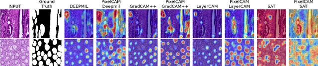

Weakly supervised object localization (WSOL) methods allow training models to classify images and localize ROIs. WSOL only requires low-cost image-class annotations yet provides a visually interpretable classifier, which is important in histology image analysis. Standard WSOL methods rely on class activation mapping (CAM) methods to produce spatial localization maps according to a single- or two-step strategy. While both strategies have made significant progress, they still face several limitations with histology images. Single-step methods can easily result in under- or over-activation due to the limited visual ROI saliency in histology images and the limited localization cues. They also face the well-known issue of asynchronous convergence between classification and localization tasks. The two-step approach is sub-optimal because it is tied to a frozen classifier, limiting the capacity for localization. Moreover, these methods also struggle when applied to out-of-distribution (OOD) datasets. In this paper, a multi-task approach for WSOL is introduced for simultaneous training of both tasks to address the asynchronous convergence problem. In particular, localization is performed in the pixel-feature space of an image encoder that is shared with classification. This allows learning discriminant features and accurate delineation of foreground/background regions to support ROI localization and image classification. We propose PixelCAM, a cost-effective foreground/background pixel-wise classifier in the pixel-feature space that allows for spatial object localization. PixelCAM is trained using pixel pseudo-labels collected from a pretrained WSOL model. Both image and pixel-wise classifiers are trained simultaneously using standard gradient descent. In addition, our pixel classifier can easily be integrated into CNN- and transformer-based architectures without any modifications.
Source-Free Domain Adaptation of Weakly-Supervised Object Localization Models for Histology
Apr 29, 2024Given the emergence of deep learning, digital pathology has gained popularity for cancer diagnosis based on histology images. Deep weakly supervised object localization (WSOL) models can be trained to classify histology images according to cancer grade and identify regions of interest (ROIs) for interpretation, using inexpensive global image-class annotations. A WSOL model initially trained on some labeled source image data can be adapted using unlabeled target data in cases of significant domain shifts caused by variations in staining, scanners, and cancer type. In this paper, we focus on source-free (unsupervised) domain adaptation (SFDA), a challenging problem where a pre-trained source model is adapted to a new target domain without using any source domain data for privacy and efficiency reasons. SFDA of WSOL models raises several challenges in histology, most notably because they are not intended to adapt for both classification and localization tasks. In this paper, 4 state-of-the-art SFDA methods, each one representative of a main SFDA family, are compared for WSOL in terms of classification and localization accuracy. They are the SFDA-Distribution Estimation, Source HypOthesis Transfer, Cross-Domain Contrastive Learning, and Adaptively Domain Statistics Alignment. Experimental results on the challenging Glas (smaller, breast cancer) and Camelyon16 (larger, colon cancer) histology datasets indicate that these SFDA methods typically perform poorly for localization after adaptation when optimized for classification.