 Add to Chrome
 Add to Firefox
 Add to Edge
Matched Outcomes, Divergent Gaze: How Foveated MLLMs Search Compared to Humans
Aug 17, 2026
Human visual search is serial: the fovea must land on a candidate to confirm it, and those landings form a scanpath. Whether multimodal large language models (MLLMs), given the same foveated input, search as humans do bears on their use as models of human vision and on attention-alignment scores. We compare three general-purpose MLLMs with human eye-movement scanpaths on goal-directed search (COCO-Search18), driving each model fixation by fixation through an identical, human-matched foveated view and assessing it along three axes: the decision of target presence, the efficiency of reaching the target, and the gaze process itself. The axes dissociate. On the decision and on target acquisition the models match or exceed humans, detecting present targets near ceiling and reaching them on the first saccade more often than people do. The gaze process is not human. Under the human-matched condition, all three share one signature: low-entropy, large-amplitude, self-consistent scanpaths that agree with themselves far more closely than two humans agree with each other. That is consistent with a single-pass, non-serial architecture rather than a limit of acuity. Matched retinal input reproduces where humans look but not how the looking unfolds in time, and no degradation regime recovers human-like search at human-like success. The gap sits on a process axis that answer-alignment and saliency metrics do not measure. Because they miss it, such metrics cannot certify human-like vision, and zero-shot models suit outcome and spatial questions but not temporal, process-level ones.

Divergent Gaze Patterns in Artistic Viewing: Spatial and Temporal Signatures of Attention Across Autistic Individuals, Artists, and Neurotypical Observers
Jul 16, 2026How different populations visually explore artworks bears on cognitive science and on accessibility design, yet most eye-tracking work in autism has used social scenes rather than art, and has analysed where the eyes land while ignoring when and in what order. We present a comparative free-viewing study across three groups, autistic adults (ASD), trained artists, and neurotypical observers, who each viewed 30 paintings for 15s. We introduce a directed, metric-grounded framework that compares groups along two complementary axes: a spatial axis, in which one group's fixation-density map predicts another's fixations under six saliency metrics (AUC-Judd, NSS, CC, SIM, KL, Information Gain); and a temporal axis, in which individual scanpaths are compared with MultiMatch, ScanMatch, a foveal-disc IoU score (FDISS), and dynamic time warping (DTW). Fixations are extracted uniformly for all groups with a dispersion-threshold algorithm. Three results converge. (i)Artists and neurotypicals are almost indistinguishable in both space (density-map correlation CC=0.96) and time (they form the most alignable scanpath pair), whereas ASD gaze diverges from both. (ii)ASD attention is dissociated: it matches artists' wide spatial exploration (dispersion, explored area) but carries a distinct temporal signature, shorter fixations, less dwell, and the most idiosyncratic (least self-consistent) scanpaths of any group. (iii)ASD gaze is not selectively artist-like on any metric; if anything it is marginally closer to neurotypical. Together these findings indicate that autistic viewing of art is a distinct, group-specific attentional profile in both space and time, and they motivate population-conditioned models of aesthetic attention. We release all analysis code and per-stimulus results.
GazeVaLM: A Multi-Observer Eye-Tracking Benchmark for Evaluating Clinical Realism in AI-Generated X-Rays
Apr 13, 2026We introduce GazeVaLM, a public eye-tracking dataset for studying clinical perception during chest radiograph authenticity assessment. The dataset comprises 960 gaze recordings from 16 expert radiologists interpreting 30 real and 30 synthetic chest X-rays (generated by diffusion based generative AI) under two conditions: diagnostic assessment and real-fake classification (Visual Turing test). For each image-observer pair, we provide raw gaze samples, fixation maps, scanpaths, saliency density maps, structured diagnostic labels, and authenticity judgments. We extend the protocol to 6 state-of-the-art multimodal LLMs, releasing their predicted diagnoses, authenticity labels, and confidence scores under matched conditions - enabling direct human-AI comparison at both decision and uncertainty levels. We further provide analyses of gaze agreement, inter-observer consistency, and benchmarking of radiologists versus LLMs in diagnostic accuracy and authenticity detection. GazeVaLM supports research in gaze modeling, clinical decision-making, human-AI comparison, generative image realism assessment, and uncertainty quantification. By jointly releasing visual attention data, clinical labels, and model predictions, we aim to facilitate reproducible research on how experts and AI systems perceive, interpret, and evaluate medical images. The dataset is available at https://huggingface.co/datasets/davidcwong/GazeVaLM.
What They Saw, Not Just Where They Looked: Semantic Scanpath Similarity via VLMs and NLP metric
Apr 09, 2026Scanpath similarity metrics are central to eye-movement research, yet existing methods predominantly evaluate spatial and temporal alignment while neglecting semantic equivalence between attended image regions. We present a semantic scanpath similarity framework that integrates vision-language models (VLMs) into eye-tracking analysis. Each fixation is encoded under controlled visual context (patch-based and marker-based strategies) and transformed into concise textual descriptions, which are aggregated into scanpath-level representations. Semantic similarity is then computed using embedding-based and lexical NLP metrics and compared against established spatial measures, including MultiMatch and DTW. Experiments on free-viewing eye-tracking data demonstrate that semantic similarity captures partially independent variance from geometric alignment, revealing cases of high content agreement despite spatial divergence. We further analyze the impact of contextual encoding on description fidelity and metric stability. Our findings suggest that multimodal foundation models enable interpretable, content-aware extensions of classical scanpath analysis, providing a complementary dimension for gaze research within the ETRA community.
SPGen: Stochastic scanpath generation for paintings using unsupervised domain adaptation
Feb 25, 2026Understanding human visual attention is key to preserving cultural heritage We introduce SPGen a novel deep learning model to predict scanpaths the sequence of eye movementswhen viewers observe paintings. Our architecture uses a Fully Convolutional Neural Network FCNN with differentiable fixation selection and learnable Gaussian priors to simulate natural viewing biases To address the domain gap between photographs and artworks we employ unsupervised domain adaptation via a gradient reversal layer allowing the model to transfer knowledge from natural scenes to paintings Furthermore a random noise sampler models the inherent stochasticity of eyetracking data. Extensive testing shows SPGen outperforms existing methods offering a powerful tool to analyze gaze behavior and advance the preservation and appreciation of artistic treasures.
Modeling Beyond MOS: Quality Assessment Models Must Integrate Context, Reasoning, and Multimodality
May 26, 2025This position paper argues that Mean Opinion Score (MOS), while historically foundational, is no longer sufficient as the sole supervisory signal for multimedia quality assessment models. MOS reduces rich, context-sensitive human judgments to a single scalar, obscuring semantic failures, user intent, and the rationale behind quality decisions. We contend that modern quality assessment models must integrate three interdependent capabilities: (1) context-awareness, to adapt evaluations to task-specific goals and viewing conditions; (2) reasoning, to produce interpretable, evidence-grounded justifications for quality judgments; and (3) multimodality, to align perceptual and semantic cues using vision-language models. We critique the limitations of current MOS-centric benchmarks and propose a roadmap for reform: richer datasets with contextual metadata and expert rationales, and new evaluation metrics that assess semantic alignment, reasoning fidelity, and contextual sensitivity. By reframing quality assessment as a contextual, explainable, and multimodal modeling task, we aim to catalyze a shift toward more robust, human-aligned, and trustworthy evaluation systems.
Shifts in Doctors' Eye Movements Between Real and AI-Generated Medical Images
Apr 21, 2025Eye-tracking analysis plays a vital role in medical imaging, providing key insights into how radiologists visually interpret and diagnose clinical cases. In this work, we first analyze radiologists' attention and agreement by measuring the distribution of various eye-movement patterns, including saccades direction, amplitude, and their joint distribution. These metrics help uncover patterns in attention allocation and diagnostic strategies. Furthermore, we investigate whether and how doctors' gaze behavior shifts when viewing authentic (Real) versus deep-learning-generated (Fake) images. To achieve this, we examine fixation bias maps, focusing on first, last, short, and longest fixations independently, along with detailed saccades patterns, to quantify differences in gaze distribution and visual saliency between authentic and synthetic images.
MoEDiff-SR: Mixture of Experts-Guided Diffusion Model for Region-Adaptive MRI Super-Resolution
Apr 09, 2025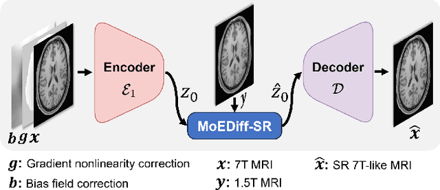
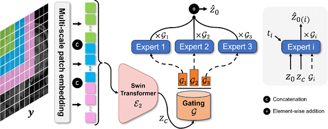
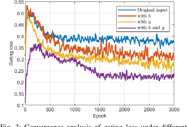
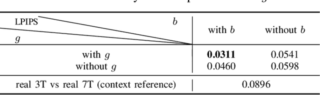
Magnetic Resonance Imaging (MRI) at lower field strengths (e.g., 3T) suffers from limited spatial resolution, making it challenging to capture fine anatomical details essential for clinical diagnosis and neuroimaging research. To overcome this limitation, we propose MoEDiff-SR, a Mixture of Experts (MoE)-guided diffusion model for region-adaptive MRI Super-Resolution (SR). Unlike conventional diffusion-based SR models that apply a uniform denoising process across the entire image, MoEDiff-SR dynamically selects specialized denoising experts at a fine-grained token level, ensuring region-specific adaptation and enhanced SR performance. Specifically, our approach first employs a Transformer-based feature extractor to compute multi-scale patch embeddings, capturing both global structural information and local texture details. The extracted feature embeddings are then fed into an MoE gating network, which assigns adaptive weights to multiple diffusion-based denoisers, each specializing in different brain MRI characteristics, such as centrum semiovale, sulcal and gyral cortex, and grey-white matter junction. The final output is produced by aggregating the denoised results from these specialized experts according to dynamically assigned gating probabilities. Experimental results demonstrate that MoEDiff-SR outperforms existing state-of-the-art methods in terms of quantitative image quality metrics, perceptual fidelity, and computational efficiency. Difference maps from each expert further highlight their distinct specializations, confirming the effective region-specific denoising capability and the interpretability of expert contributions. Additionally, clinical evaluation validates its superior diagnostic capability in identifying subtle pathological features, emphasizing its practical relevance in clinical neuroimaging. Our code is available at https://github.com/ZWang78/MoEDiff-SR.
Eyes Tell the Truth: GazeVal Highlights Shortcomings of Generative AI in Medical Imaging
Mar 26, 2025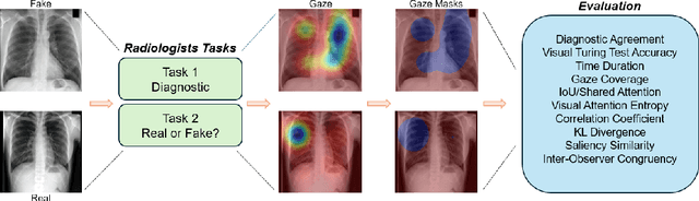
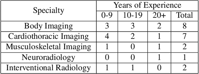

The demand for high-quality synthetic data for model training and augmentation has never been greater in medical imaging. However, current evaluations predominantly rely on computational metrics that fail to align with human expert recognition. This leads to synthetic images that may appear realistic numerically but lack clinical authenticity, posing significant challenges in ensuring the reliability and effectiveness of AI-driven medical tools. To address this gap, we introduce GazeVal, a practical framework that synergizes expert eye-tracking data with direct radiological evaluations to assess the quality of synthetic medical images. GazeVal leverages gaze patterns of radiologists as they provide a deeper understanding of how experts perceive and interact with synthetic data in different tasks (i.e., diagnostic or Turing tests). Experiments with sixteen radiologists revealed that 96.6% of the generated images (by the most recent state-of-the-art AI algorithm) were identified as fake, demonstrating the limitations of generative AI in producing clinically accurate images.
Feasibility study for reconstruction of knee MRI from one corresponding X-ray via CNN
Mar 16, 2025Generally, X-ray, as an inexpensive and popular medical imaging technique, is widely chosen by medical practitioners. With the development of medical technology, Magnetic Resonance Imaging (MRI), an advanced medical imaging technique, has already become a supplementary diagnostic option for the diagnosis of KOA. We propose in this paper a deep-learning-based approach for generating MRI from one corresponding X-ray. Our method uses the hidden variables of a Convolutional Auto-Encoder (CAE) model, trained for reconstructing X-ray image, as inputs of a generator model to provide 3D MRI.