 Add to Chrome
 Add to Firefox
 Add to Edge
Configurable γ Photon Spectrometer to Enable Precision Radioguided Tumor Resection
Dec 16, 2025Surgical tumor resection aims to remove all cancer cells in the tumor margin and at centimeter-scale depths below the tissue surface. During surgery, microscopic clusters of disease are intraoperatively difficult to visualize and are often left behind, significantly increasing the risk of cancer recurrence. Radioguided surgery (RGS) has shown the ability to selectively tag cancer cells with gamma (γ) photon emitting radioisotopes to identify them, but require a mm-scale γ photon spectrometer to localize the position of these cells in the tissue margin (i.e., a function of incident γ photon energy) with high specificity. Here we present a 9.9 mm2 integrated circuit (IC)-based γ spectrometer implemented in 180 nm CMOS, to enable the measurement of single γ photons and their incident energy with sub-keV energy resolution. We use small 2 2 um reverse-biased diodes that have low depletion region capacitance, and therefore produce millivolt-scale voltage signals in response to the small charge generated by incident γ photons. A low-power energy spectrometry method is implemented by measuring the decay time it takes for the generated voltage signal to settle back to DC after a γ detection event, instead of measuring the voltage drop directly. This spectrometry method is implemented in three different pixel architectures that allow for configurable pixel sensitivity, energy-resolution, and energy dynamic range based on the widely heterogenous surgical and patient presentation in RGS. The spectrometer was tested with three common γ-emitting radioisotopes (64Cu, 133Ba, 177Lu), and is able to resolve activities down to 1 uCi with sub-keV energy resolution and 1.315 MeV energy dynamic range, using 5-minute acquisitions.
Joint Correction of Attenuation and Scatter Using Deep Convolutional Neural Networks (DCNN) for Time-of-Flight PET
Nov 28, 2018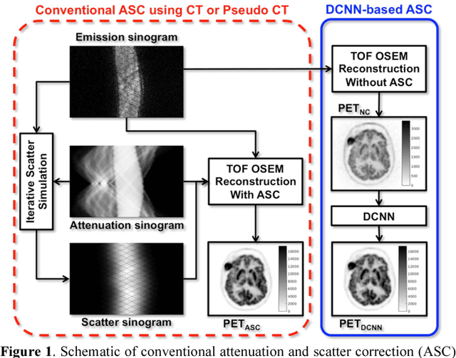
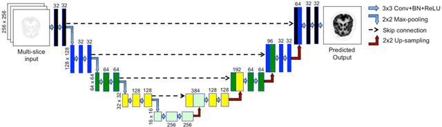
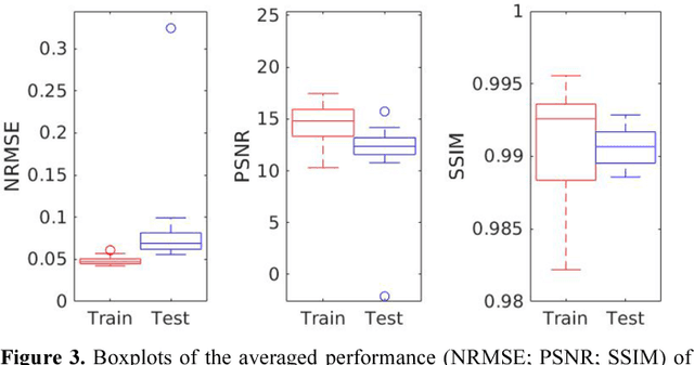
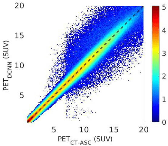
Deep convolutional neural networks (DCNN) have demonstrated its capability to convert MR image to pseudo CT for PET attenuation correction in PET/MRI. Conventionally, attenuated events are corrected in sinogram space using attenuation maps derived from CT or MR-derived pseudo CT. Separately, scattered events are iteratively estimated by a 3D model-based simulation using down-sampled attenuation and emission sinograms. However, no studies have investigated joint correction of attenuation and scatter using DCNN in image space. Therefore, we aim to develop and optimize a DCNN model for attenuation and scatter correction (ASC) simultaneously in PET image space without additional anatomical imaging or time-consuming iterative scatter simulation. For the first time, we demonstrated the feasibility of directly producing PET images corrected for attenuation and scatter using DCNN (PET-DCNN) from noncorrected PET (PET-NC) images.
Attenuation correction for brain PET imaging using deep neural network based on dixon and ZTE MR images
May 24, 2018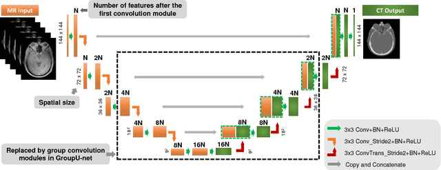
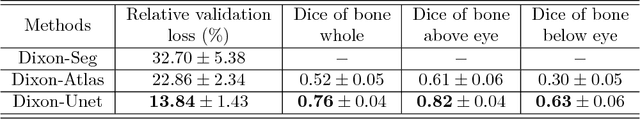
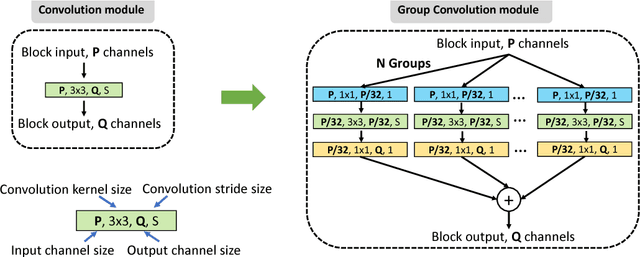
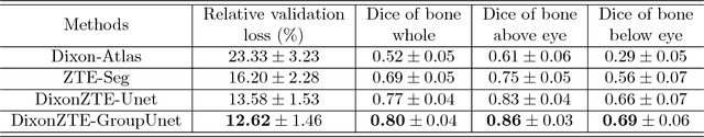
Positron Emission Tomography (PET) is a functional imaging modality widely used in neuroscience studies. To obtain meaningful quantitative results from PET images, attenuation correction is necessary during image reconstruction. For PET/MR hybrid systems, PET attenuation is challenging as Magnetic Resonance (MR) images do not reflect attenuation coefficients directly. To address this issue, we present deep neural network methods to derive the continuous attenuation coefficients for brain PET imaging from MR images. With only Dixon MR images as the network input, the existing U-net structure was adopted and analysis using forty patient data sets shows it is superior than other Dixon based methods. When both Dixon and zero echo time (ZTE) images are available, we have proposed a modified U-net structure, named GroupU-net, to efficiently make use of both Dixon and ZTE information through group convolution modules when the network goes deeper. Quantitative analysis based on fourteen real patient data sets demonstrates that both network approaches can perform better than the standard methods, and the proposed network structure can further reduce the PET quantification error compared to the U-net structure.