 Add to Chrome
 Add to Firefox
 Add to Edge
Simultaneous Polysomnography and Cardiotocography Reveal Temporal Correlation Between Maternal Obstructive Sleep Apnea and Fetal Hypoxia
Apr 17, 2025Background: Obstructive sleep apnea syndrome (OSAS) during pregnancy is common and can negatively affect fetal outcomes. However, studies on the immediate effects of maternal hypoxia on fetal heart rate (FHR) changes are lacking. Methods: We used time-synchronized polysomnography (PSG) and cardiotocography (CTG) data from two cohorts to analyze the correlation between maternal hypoxia and FHR changes (accelerations or decelerations). Maternal hypoxic event characteristics were analyzed using generalized linear modeling (GLM) to assess their associations with different FHR changes. Results: A total of 118 pregnant women participated. FHR changes were significantly associated with maternal hypoxia, primarily characterized by accelerations. A longer hypoxic duration correlated with more significant FHR accelerations (P < 0.05), while prolonged hypoxia and greater SpO2 drop were linked to FHR decelerations (P < 0.05). Both cohorts showed a transient increase in FHR during maternal hypoxia, which returned to baseline after the event resolved. Conclusion: Maternal hypoxia significantly affects FHR, suggesting that maternal OSAS may contribute to fetal hypoxia. These findings highlight the importance of maternal-fetal interactions and provide insights for future interventions.
A Computer-Aided Diagnosis System for Breast Pathology: A Deep Learning Approach with Model Interpretability from Pathological Perspective
Aug 05, 2021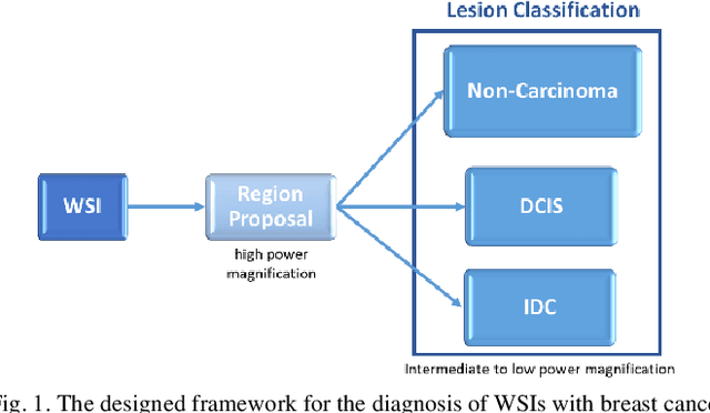
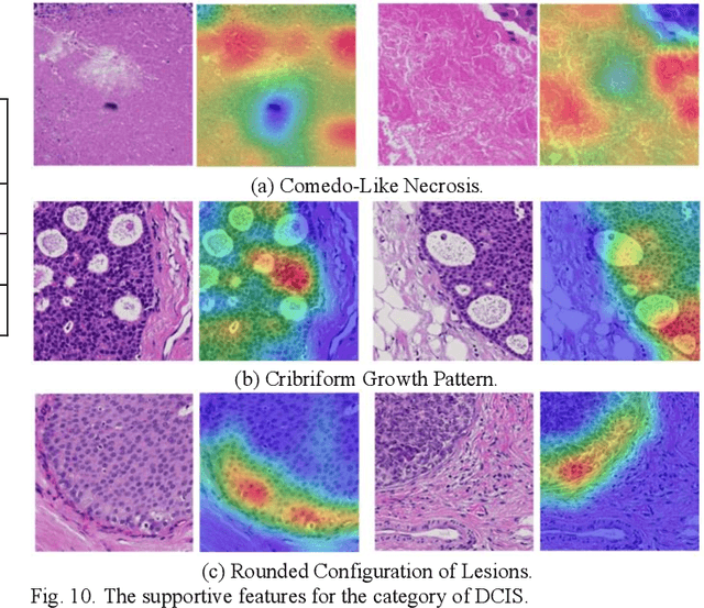
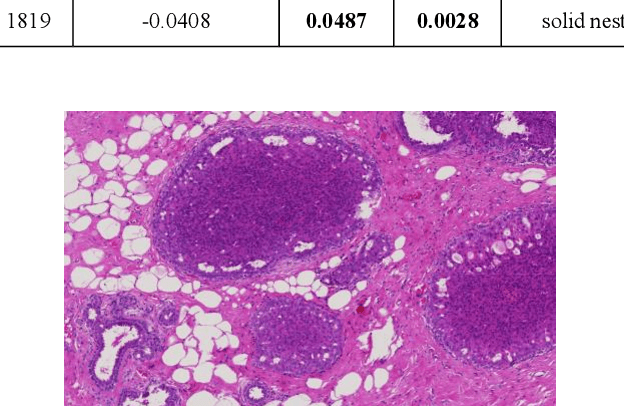
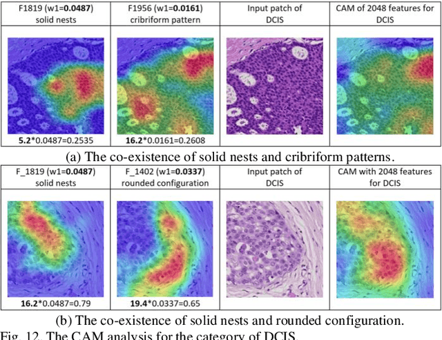
Objective: We develop a computer-aided diagnosis (CAD) system using deep learning approaches for lesion detection and classification on whole-slide images (WSIs) with breast cancer. The deep features being distinguishing in classification from the convolutional neural networks (CNN) are demonstrated in this study to provide comprehensive interpretability for the proposed CAD system using pathological knowledge. Methods: In the experiment, a total of 186 slides of WSIs were collected and classified into three categories: Non-Carcinoma, Ductal Carcinoma in Situ (DCIS), and Invasive Ductal Carcinoma (IDC). Instead of conducting pixel-wise classification into three classes directly, we designed a hierarchical framework with the multi-view scheme that performs lesion detection for region proposal at higher magnification first and then conducts lesion classification at lower magnification for each detected lesion. Results: The slide-level accuracy rate for three-category classification reaches 90.8% (99/109) through 5-fold cross-validation and achieves 94.8% (73/77) on the testing set. The experimental results show that the morphological characteristics and co-occurrence properties learned by the deep learning models for lesion classification are accordant with the clinical rules in diagnosis. Conclusion: The pathological interpretability of the deep features not only enhances the reliability of the proposed CAD system to gain acceptance from medical specialists, but also facilitates the development of deep learning frameworks for various tasks in pathology. Significance: This paper presents a CAD system for pathological image analysis, which fills the clinical requirements and can be accepted by medical specialists with providing its interpretability from the pathological perspective.
Understanding the Mechanism of Deep Learning Framework for Lesion Detection in Pathological Images with Breast Cancer
Mar 04, 2019


The computer-aided detection (CADe) systems are developed to assist pathologists in slide assessment, increasing diagnosis efficiency and reducing missing inspections. Many studies have shown such a CADe system with deep learning approaches outperforms the one using conventional methods that rely on hand-crafted features based on field-knowledge. However, most developers who adopted deep learning models directly focused on the efficacy of outcomes, without providing comprehensive explanations on why their proposed frameworks can work effectively. In this study, we designed four experiments to verify the consecutive concepts, showing that the deep features learned from pathological patches are interpretable by domain knowledge of pathology and enlightening for clinical diagnosis in the task of lesion detection. The experimental results show the activation features work as morphological descriptors for specific cells or tissues, which agree with the clinical rules in classification. That is, the deep learning framework not only detects the distribution of tumor cells but also recognizes lymphocytes, collagen fibers, and some other non-cell structural tissues. Most of the characteristics learned by the deep learning models have summarized the detection rules that can be recognized by the experienced pathologists, whereas there are still some features may not be intuitive to domain experts but discriminative in classification for machines. Those features are worthy to be further studied in order to find out the reasonable correlations to pathological knowledge, from which pathological experts may draw inspirations for exploring new characteristics in diagnosis.