 Add to Chrome
 Add to Firefox
 Add to Edge
PC-Seg: Progressive Cross-View Consistency for 3D OCT Segmentation from Sparse 2D Annotations
Jul 21, 2026
Volumetric segmentation of optical coherence tomography (OCT) images is essential for diagnosing ocular diseases but requires labor-intensive voxel-wise annotations. While semi-supervised learning (SSL) can reduce annotation costs, most existing methods process data slice by slice and fail to exploit the inherent 3D spatial context. We propose PC-Seg, a progressive cross-view consistency framework that learns high-accuracy 3D segmentation models from sparse 2D annotations. Unlike conventional multi-view approaches, PC-Seg uses a single 2D model to learn cross-view consistency from standard B-scans and orthogonal slices, thereby generating reliable volumetric pseudo-labels. These pseudo-labels are then distilled into a 3D model, followed by a co-training stage in which the 2D and 3D models mutually refine each other through ensemble pseudo-labeling. Experiments on the MSHC and Duke DME datasets demonstrate that PC-Seg achieves accuracy comparable to fully supervised learning while using labels for only about 0.7% of the training data, outperforming state-of-the-art semi-supervised and retinal layer segmentation methods. Our code is publicly available at https://github.com/gsisaoki/pc-seg-official.

Formula-Driven Data Augmentation and Partial Retinal Layer Copying for Retinal Layer Segmentation
Oct 02, 2024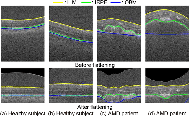

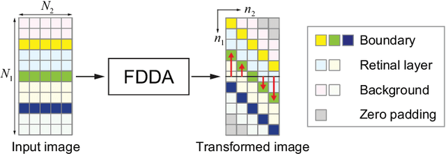
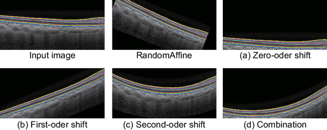
Major retinal layer segmentation methods from OCT images assume that the retina is flattened in advance, and thus cannot always deal with retinas that have changes in retinal structure due to ophthalmopathy and/or curvature due to myopia. To eliminate the use of flattening in retinal layer segmentation for practicality of such methods, we propose novel data augmentation methods for OCT images. Formula-driven data augmentation (FDDA) emulates a variety of retinal structures by vertically shifting each column of the OCT images according to a given mathematical formula. We also propose partial retinal layer copying (PRLC) that copies a part of the retinal layers and pastes it into a region outside the retinal layers. Through experiments using the OCT MS and Healthy Control dataset and the Duke Cyst DME dataset, we demonstrate that the use of FDDA and PRLC makes it possible to detect the boundaries of retinal layers without flattening even retinal layer segmentation methods that assume flattening of the retina.