 Add to Chrome
 Add to Firefox
 Add to Edge
Quality assessment of brain structural MR images: Comparing generalization of deep learning versus hand-crafted feature-based machine learning methods to new sites
Mar 17, 2026Quality assessment of brain structural MR images is critical for large-scale neuroimaging studies, where motion artifacts can significantly bias clinical estimates. While visual rating remains the gold standard, it is time-consuming and subjective. This study evaluates the relative performance and generalization capabilities of two prominent Automated Quality Assessment (AQA) methods: MRIQC, which uses hand-crafted image-quality metrics with traditional machine learning, and CNNQC, which utilizes a deep learning (DL) architecture. Using a heterogeneous dataset of 1,098 T1-weighted volumes from 17 different sites, we assessed performance on both seen sites and entirely new sites using a leave-one-site-out (LOSO) approach. Our results indicate that both DL and traditional ML methods struggle to generalize to new scanners or sites. While MRIQC generally achieved higher accuracy across most unseen sites, CNNQC demonstrated higher sensitivity for detecting poor-quality scans. Given that DL-based methods like CNNQC offer higher computational efficiency and do not require expensive pre-processing, they may be preferred for widespread deployment, provided that future work focuses on improving cross-site generalizability.
Using convolution neural networks to learn enhanced fiber orientation distribution models from commercially available diffusion magnetic resonance imaging
Aug 12, 2020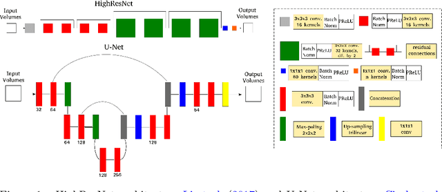
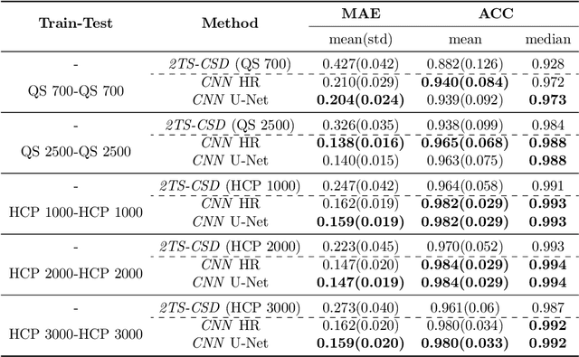
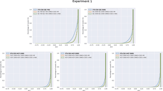
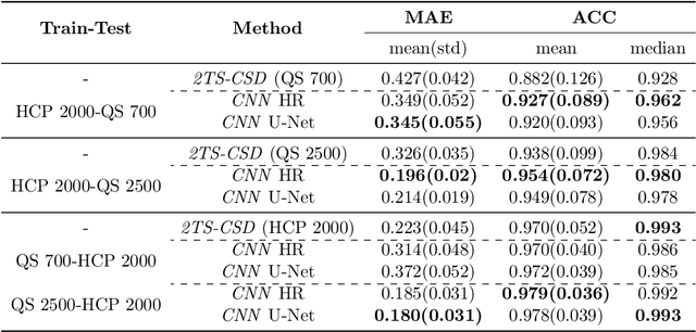
Accurate local fiber orientation distribution (FOD) modeling based on diffusion magnetic resonance imaging (dMRI) capable of resolving complex fiber configurations benefit from specific acquisition protocols that impose a high number of gradient directions (b-vecs), a high maximum b-value (b-vals) and multiple b-values (multi-shell). However, acquisition time is limited in a clinical setting and commercial scanners may not provide robust state-of-the-art dMRI sequences. Therefore, dMRI is often acquired as single-shell (SS) (single b-value). Here, we learn improved FODs for commercially acquired dMRI. We evaluate the use of 3D convolutional neural networks (CNNs) to regress multi-shell FOS representations from single-shell representations, using the spherical harmonics basis obtained from constrained spherical deconvolution (CSD) to model FODs. We use U-Net and HighResNet 3D CNN architectures and data from the publicly available Human Connectome Dataset and a dataset acquired at National Hospital For Neurology and Neurosurgery Queen Square. We evaluate how well the CNN models can resolve local fiber orientation 1) when training and testing on datasets with same dMRI acquisition protocol; 2) when testing on dataset with a different dMRI acquisition protocol than used training the CNN models; and 3) when testing on datasets with a fewer number dMRI gradient directions than used training the CNN models. Our approach may enable robust CSD model estimation on dMRI acquisition protocols which are single shell and with a few gradient directions, reducing acquisition times, and thus, facilitating translation to time-limited clinical environments.