 Add to Chrome
 Add to Firefox
 Add to Edge
SMILE-UHURA Challenge -- Small Vessel Segmentation at Mesoscopic Scale from Ultra-High Resolution 7T Magnetic Resonance Angiograms
Nov 14, 2024The human brain receives nutrients and oxygen through an intricate network of blood vessels. Pathology affecting small vessels, at the mesoscopic scale, represents a critical vulnerability within the cerebral blood supply and can lead to severe conditions, such as Cerebral Small Vessel Diseases. The advent of 7 Tesla MRI systems has enabled the acquisition of higher spatial resolution images, making it possible to visualise such vessels in the brain. However, the lack of publicly available annotated datasets has impeded the development of robust, machine learning-driven segmentation algorithms. To address this, the SMILE-UHURA challenge was organised. This challenge, held in conjunction with the ISBI 2023, in Cartagena de Indias, Colombia, aimed to provide a platform for researchers working on related topics. The SMILE-UHURA challenge addresses the gap in publicly available annotated datasets by providing an annotated dataset of Time-of-Flight angiography acquired with 7T MRI. This dataset was created through a combination of automated pre-segmentation and extensive manual refinement. In this manuscript, sixteen submitted methods and two baseline methods are compared both quantitatively and qualitatively on two different datasets: held-out test MRAs from the same dataset as the training data (with labels kept secret) and a separate 7T ToF MRA dataset where both input volumes and labels are kept secret. The results demonstrate that most of the submitted deep learning methods, trained on the provided training dataset, achieved reliable segmentation performance. Dice scores reached up to 0.838 $\pm$ 0.066 and 0.716 $\pm$ 0.125 on the respective datasets, with an average performance of up to 0.804 $\pm$ 0.15.
Fourier PD and PDUNet: Complex-valued networks to speed-up MR Thermometry during Hypterthermia
Oct 02, 2023Hyperthermia (HT) in combination with radio- and/or chemotherapy has become an accepted cancer treatment for distinct solid tumour entities. In HT, tumour tissue is exogenously heated to temperatures of 39 to 43 $\degree$C for 60 minutes. Temperature monitoring can be performed noninvasively using dynamic magnetic resonance imaging (MRI). However, the slow nature of MRI leads to motion artefacts in the images due to the movements of patients during image acquisition time. By discarding parts of the data, the speed of the acquisition can be increased - known as Undersampling. However, due to the invalidation of the Nyquist criterion, the acquired images have lower resolution and can also produce artefacts. The aim of this work was, therefore, to reconstruct highly undersampled MR thermometry acquisitions with better resolution and with less artefacts compared to conventional techniques like compressed sensing. The use of deep learning in the medical field has emerged in recent times, and various studies have shown that deep learning has the potential to solve inverse problems such as MR image reconstruction. However, most of the published work only focusses on the magnitude images, while the phase images are ignored, which are fundamental requirements for MR thermometry. This work, for the first time ever, presents deep learning based solutions for reconstructing undersampled MR thermometry data. Two different deep learning models have been employed here, the Fourier Primal-Dual network and Fourier Primal-Dual UNet, to reconstruct highly undersampled complex images of MR thermometry. It was observed that the method was able to reduce the temperature difference between the undersampled MRIs and the fully sampled MRIs from 1.5 $\degree$C to 0.5 $\degree$C.
Exploration of Interpretability Techniques for Deep COVID-19 Classification using Chest X-ray Images
Jun 10, 2020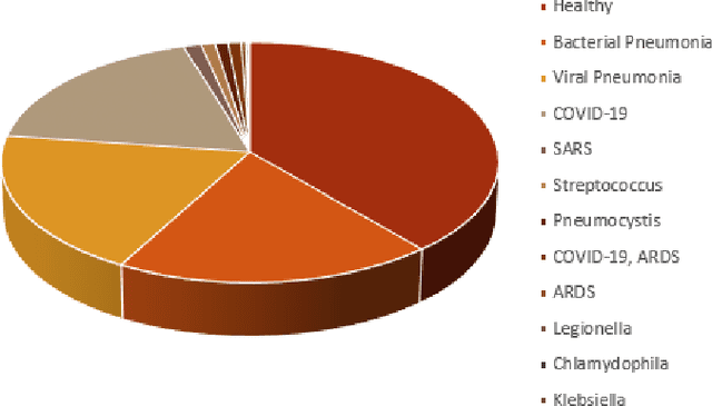
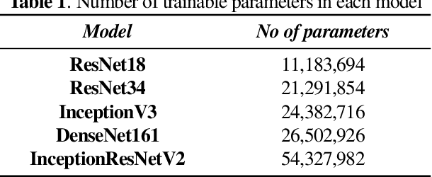
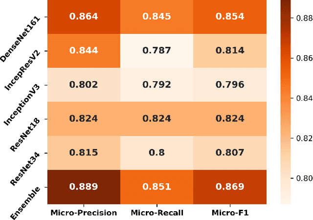
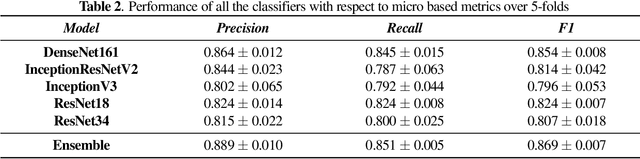
The outbreak of COVID-19 has shocked the entire world with its fairly rapid spread and has challenged different sectors. One of the most effective ways to limit its spread is the early and accurate diagnosis of infected patients. Medical imaging such as X-ray and Computed Tomography (CT) combined with the potential of Artificial Intelligence (AI) plays an essential role in supporting the medical staff in the diagnosis process. Thereby, the use of five different deep learning models (ResNet18, ResNet34, InceptionV3, InceptionResNetV2, and DenseNet161) and their Ensemble have been used in this paper, to classify COVID-19, pneumoni{\ae} and healthy subjects using Chest X-Ray. Multi-label classification was performed to predict multiple pathologies for each patient, if present. Foremost, the interpretability of each of the networks was thoroughly studied using techniques like occlusion, saliency, input X gradient, guided backpropagation, integrated gradients, and DeepLIFT. The mean Micro-F1 score of the models for COVID-19 classifications ranges from 0.66 to 0.875, and is 0.89 for the Ensemble of the network models. The qualitative results depicted the ResNets to be the most interpretable model.
Machine learning approach for segmenting glands in colon histology images using local intensity and texture features
May 15, 2019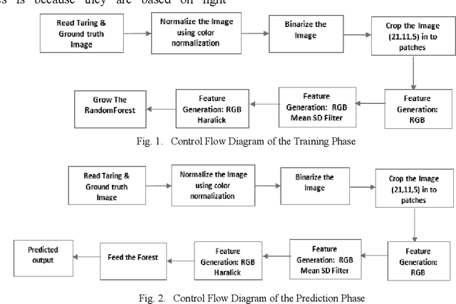
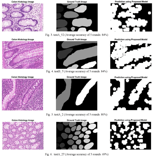
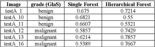
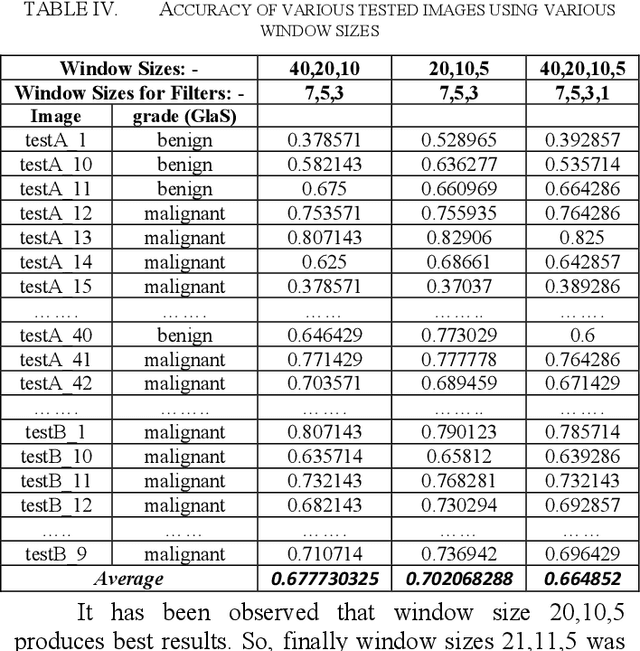
Colon Cancer is one of the most common types of cancer. The treatment is planned to depend on the grade or stage of cancer. One of the preconditions for grading of colon cancer is to segment the glandular structures of tissues. Manual segmentation method is very time-consuming, and it leads to life risk for the patients. The principal objective of this project is to assist the pathologist to accurate detection of colon cancer. In this paper, the authors have proposed an algorithm for an automatic segmentation of glands in colon histology using local intensity and texture features. Here the dataset images are cropped into patches with different window sizes and taken the intensity of those patches, and also calculated texture-based features. Random forest classifier has been used to classify this patch into different labels. A multilevel random forest technique in a hierarchical way is proposed. This solution is fast, accurate and it is very much applicable in a clinical setup.