 Add to Chrome
 Add to Firefox
 Add to Edge
AlcLaM: Arabic Dialectal Language Model
Jul 18, 2024

Pre-trained Language Models (PLMs) are integral to many modern natural language processing (NLP) systems. Although multilingual models cover a wide range of languages, they often grapple with challenges like high inference costs and a lack of diverse non-English training data. Arabic-specific PLMs are trained predominantly on modern standard Arabic, which compromises their performance on regional dialects. To tackle this, we construct an Arabic dialectal corpus comprising 3.4M sentences gathered from social media platforms. We utilize this corpus to expand the vocabulary and retrain a BERT-based model from scratch. Named AlcLaM, our model was trained using only 13 GB of text, which represents a fraction of the data used by existing models such as CAMeL, MARBERT, and ArBERT, compared to 7.8%, 10.2%, and 21.3%, respectively. Remarkably, AlcLaM demonstrates superior performance on a variety of Arabic NLP tasks despite the limited training data. AlcLaM is available at GitHub https://github.com/amurtadha/Alclam and HuggingFace https://huggingface.co/rahbi.
Brain tumor multi classification and segmentation in MRO images using deep learning
Apr 20, 2023

This study proposes a deep learning model for the classification and segmentation of brain tumors from magnetic resonance imaging (MRI) scans. The classification model is based on the EfficientNetB1 architecture and is trained to classify images into four classes: meningioma, glioma, pituitary adenoma, and no tumor. The segmentation model is based on the U-Net architecture and is trained to accurately segment the tumor from the MRI images. The models are evaluated on a publicly available dataset and achieve high accuracy and segmentation metrics, indicating their potential for clinical use in the diagnosis and treatment of brain tumors.
Brain Tumor classification and Segmentation using Deep Learning
Apr 16, 2023
Brain tumors are a complex and potentially life-threatening medical condition that requires accurate diagnosis and timely treatment. In this paper, we present a machine learning-based system designed to assist healthcare professionals in the classification and diagnosis of brain tumors using MRI images. Our system provides a secure login, where doctors can upload or take a photo of MRI and our app can classify the model and segment the tumor, providing the doctor with a folder of each patient's history, name, and results. Our system can also add results or MRI to this folder, draw on the MRI to send it to another doctor, and save important results in a saved page in the app. Furthermore, our system can classify in less than 1 second and allow doctors to chat with a community of brain tumor doctors. To achieve these objectives, our system uses a state-of-the-art machine learning algorithm that has been trained on a large dataset of MRI images. The algorithm can accurately classify different types of brain tumors and provide doctors with detailed information on the size, location, and severity of the tumor. Additionally, our system has several features to ensure its security and privacy, including secure login and data encryption. We evaluated our system using a dataset of real-world MRI images and compared its performance to other existing systems. Our results demonstrate that our system is highly accurate, efficient, and easy to use. We believe that our system has the potential to revolutionize the field of brain tumor diagnosis and treatment and provide healthcare professionals with a powerful tool for improving patient outcomes.
An ENAS Based Approach for Constructing Deep Learning Models for Breast Cancer Recognition from Ultrasound Images
May 27, 2020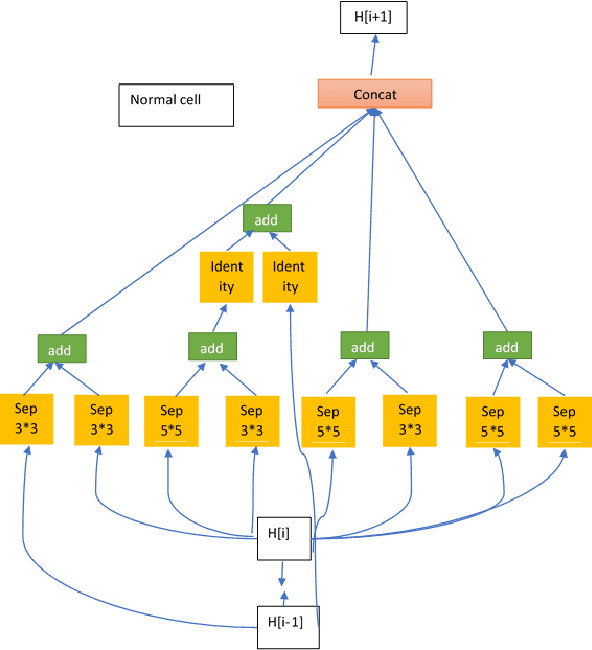
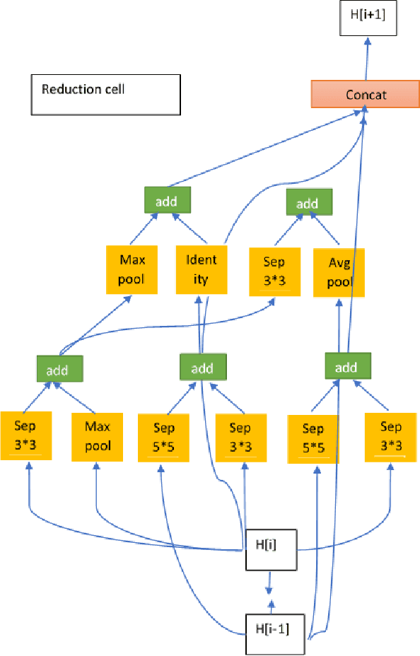
Deep Convolutional Neural Networks (CNN) provides an "end-to-end" solution for image pattern recognition with impressive performance in many areas of application including medical imaging. Most CNN models of high performance use hand-crafted network architectures that require expertise in CNNs to utilise their potentials. In this paper, we applied the Efficient Neural Architecture Search (ENAS) method to find optimal CNN architectures for classifying breast lesions from ultrasound (US) images. Our empirical study with a dataset of 524 US images shows that the optimal models generated by using ENAS achieve an average accuracy of 89.3%, surpassing other hand-crafted alternatives. Furthermore, the models are simpler in complexity and more efficient. Our study demonstrates that the ENAS approach to CNN model design is a promising direction for classifying ultrasound images of breast lesions.